Friday, June 20, 2025

Doublet Design Overcomes a Metalens Bottleneck in Microscopy


A research team from Nanjing University has developed a solution that improves metalens performance in and for microscopy applications. The researchers’ metalens-based microscope achieves both a wide field of view (FOV) and high-resolution imaging, addressing the inherent trade-off between these two critical parameters that, according to the researchers, has prevented metalenses to date from achieving performance comparable to conventional microscopes.

Metalenses face significant challenges in practical microscopy applications. Off-axis aberrations, which severely restrict metalens FOV and resolution capabilities, are the primary limitations to the use of metalenses in practical microscopy.

In their system architecture, the researchers used a doublet configuration of two metalenses on opposite sides of a transparent silica substrate combined with annular illumination. The two metalenses consist of silicon nitride nano-fins, crafted as high-aspect-ratio squares with precise dimensions and arranged at carefully calculated intervals. This design approach mitigates off-axis aberrations and increases resolution capabilities, thereby optimizing imaging performance.

The researchers’ prototype had a 1-mm FOV with a half-pitch resolution of 620 nm. Additionally, the prototype is compact, with a measurement of 4 cm × 4 cm × 5 cm. The researchers used their meta-microscope prototype to image cervical cancer cells. The system captured images of various stages of cancer development within the same FOV, revealing important cellular details such as nuclear enlargement, deformation, and division.

“Our experimental results demonstrate high-quality microscopic bioimages that are comparable to those obtained from traditional microscopes within a compact prototype, highlighting its potential applications in portable and convenient settings,” said Li.

According to the researchers, the technology could be used for research scenarios where traditional microscopes would be impractical. Further, it could be integrated into microelectronic devices and systems for clinical or biomedical automation.

Bio Photonics Research Award


Visit: biophotonicsresearch.com
Nominate Now: https://biophotonicsresearch.com/award-nomination/?ecategory=Awards&rcategory=Awardee

#MeatAnalysis #FluorescenceTech #FoodQuality #FoodSafety #SpectroscopyInFood #MeatAuthentication #RapidDetection #FoodScience #MeatFreshness #MolecularDetection #FoodIndustryInnovation #NonDestructiveTesting #FoodMonitoring #SpectroscopyApplications #QualityControl #AdvancedSpectroscopy #MeatSpoilageDetection #FoodIntegrity #SmartFoodTesting #RealTimeAnalysis #FoodAuthenticity #FoodSafetyInnovation #SpectroscopyResearch #NextGenFoodSafety #InnovativeFoodScience,

Thursday, June 19, 2025

Raman Spectroscopy Undergoes Major Speed Upgrade


Researchers at the University of Tokyo have increased the measurement rate of Raman spectroscopy by 100-fold. Since the measurement rate of the technique has been a major limitation, the improvement is expected to aid advancements in multiple fields relying on the identification of molecules and cells, such as biomedical diagnostics and material analytics.

As a mode of identification for cells and molecules, Raman spectroscopy is widely used, but it’s limited in its ability to keep up with the speed of changes in certain chemical and physical reactions due to the low scattering cross section.

Over the last decade, various broadband-coherent Raman scattering spectroscopy techniques have been developed to address the limitation, achieving a measurement of 500 kSpectra/s (kilospectra per second).

In order to further improve the measurement rate, the team built a system from scratch, leveraging a mode-locked ytterbium laser system developed by Takuro Ideguchi and his team at the Institute for Photon Science and Technology at the University of Tokyo.

In building the system, the team combined coherent Raman spectroscopy — a version of Raman spectroscopy that produces stronger signals than the conventional, spontaneous Raman spectroscopy— with their previously developed specifically designed ultrashort pulse laser and time-stretch technology using optical fibers.

The developed system provides a 50 MSpectra/s (megaspectra per second) measurement rate, a 100-fold increase compared to the previous fastest rate of 500 kSpectra/s. The system enables highly efficient Raman scattering with an ultrashort femtosecond pulse and sensitive time-stretch detection with picosecond probe pulse at a high repetition of the laser.

As a proof-of-concept, the team measured broadband coherent Stokes Raman scattering spectra of organic compounds covering the molecular fingerprint region from 200 to 1200 cm-1.

“We aim to apply our spectrometer to microscopy, enabling the capture of 2D or 3D images with Raman scattering spectra,” Ideguchi said. “Additionally, we envision its use in flow cytometry by combining this technology with microfluidics. These systems will enable high-throughput, label-free chemical imaging and spectroscopy of biomolecules in cells or tissues.”

According to the researchers, the high-speed broadband vibrational spectroscopy technique holds promise for unprecedented measurements of sub-microsecond dynamics of irreversible phenomena and extremely high throughput measurements.

Bio Photonics Research Award

Visit: biophotonicsresearch.com
Nominate Now: https://biophotonicsresearch.com/award-nomination/?ecategory=Awards&rcategory=Awardee

#MeatAnalysis #FluorescenceTech #FoodQuality #FoodSafety #SpectroscopyInFood #MeatAuthentication #RapidDetection #FoodScience #MeatFreshness #MolecularDetection #FoodIndustryInnovation #NonDestructiveTesting #FoodMonitoring #SpectroscopyApplications #QualityControl #AdvancedSpectroscopy #MeatSpoilageDetection #FoodIntegrity #SmartFoodTesting #RealTimeAnalysis #FoodAuthenticity #FoodSafetyInnovation #SpectroscopyResearch #NextGenFoodSafety #InnovativeFoodScience,
Wednesday, June 18, 2025

Light-Based Control of Bacteria Aims to Quell Antibiotic Resistance


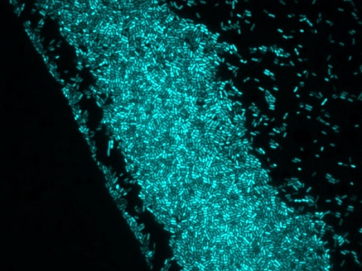
Bacterial resistance to antibiotics is a growing challenge for the healthcare and environmental sectors. Bacterial persistence is usually the first step leading to resistance, which involves a change in an organism’s genome.

In the future, it may be possible to address antibiotic persistence and resistance by using light to regulate bacterial response to antibiotics. A technique developed at the Polytechnic University of Milan (Politecnico di Milano), by a team participating in the Engineering of Bacteria to See Light (EOS) project, allows bacteria to sense light and convert light energy into electrical signals across the bacterial membrane, without the need for genetic modification.

To demonstrate photocontrol of bacterial membrane potential, the researchers attached phototransducing molecules to bacterial surfaces to make the bacteria light-responsive. When the researchers exposed the phototransducers to light, the electrical potential of the bacterial membrane changed.

The researchers used Ziapin2, a photoswitchable chemical compound, with two broad-spectrum antibiotics, Kanamycin and Ampicillin. These antibiotics have different modes of activation. Kanamycin requires cytosolic uptake to inhibit protein synthesis, while Ampicillin targets cell wall polymerization at the cell envelope.

When the researchers exposed B. subtilis to Kanamycin and Ziapin2 through photoactivation with 470-nm light, the previously lethal effects of Kanamycin were markedly reduced. In contrast, Ampicillin-treated samples remained largely unaffected by light-induced membrane modulation.

“This interplay between light and electrical signaling allows us to control key biological processes such as movement, biofilm formation, and antibiotic sensitivity,” professor Giuseppe Maria Paternò said. “By manipulating the membrane potential on demand, we can influence antibiotic uptake and restore or even enhance the effectiveness of treatments against resistant strains.”

Possible applications for the technique include developing next-generation antimicrobial platforms that use light to target resistant pathogens, and biocompatible, light-guided “bacterial robots” capable of delivering drugs to specific areas of the body — even the areas that are hard to reach, such as the gastrointestinal tract.

More broadly, the potential to modulate and control the interaction between bacterial cells and antibiotics using light and synthetic photoswitches, without genetic engineering, in real time, represents a first step in developing a new approach to combating antibiotic resistance.

In the future, photoswitchable chemical compounds could be membrane-targeted and tailored to work with different classes of antibiotics, advancing bacterial bioelectric regulation and its applications in antimicrobial therapies.


Bio Photonics Research Award

Visit: biophotonicsresearch.com
Nominate Now: https://biophotonicsresearch.com/award-nomination/?ecategory=Awards&rcategory=Awardee

#MeatAnalysis #FluorescenceTech #FoodQuality #FoodSafety #SpectroscopyInFood #MeatAuthentication #RapidDetection #FoodScience #MeatFreshness #MolecularDetection #FoodIndustryInnovation #NonDestructiveTesting #FoodMonitoring #SpectroscopyApplications #QualityControl #AdvancedSpectroscopy #MeatSpoilageDetection #FoodIntegrity #SmartFoodTesting #RealTimeAnalysis #FoodAuthenticity #FoodSafetyInnovation #SpectroscopyResearch #NextGenFoodSafety #InnovativeFoodScience,

Tuesday, June 17, 2025

Advanced OCT System Integral to Eye Transplantation


An adaptation of OCT put into use at Northwestern University, visible OCT (vis-OCT), will be a key component in an Advanced Research Projects Agency for Health (ARPA-H)-funded project that aims to conduct successful eye transplants in the next several years. The technology will be used to examine the structural integrity of eyes in donors, such as the health of retinal layers and the flow of blood as well as monitoring their continued viability throughout the process.

This work will be supported by over $50 million in ARPA-H funds to multiple institutions, earmarked for viability, imaging, surgical, immunomodulation, ocular preservation, and neuroregeneration strategies within the Transplantation of Human Eye Allografts (THEA) project. The Northwestern team, led by Hao Zhang, a professor of biomedical engineering, and Cheng Sun, a professor of mechanical engineering, will be part of group directed by Jeffrey Goldberg, professor and chair of ophthalmology at the Byers Eye Institute at Stanford University.

Opticent Health, a spinoff company of Zhang’s lab at Northwestern, created the Halo 375 for animal research and the Aurora X4 for human research. These center on a vis-OCT system that includes a supercontinuum white light laser based on photonic crystals, fundus camera for fast imaging, an adjustable field of view, and software to analyze the generated data. It has not yet been approved by the FDA, but is in use for clinical research.

“At this point, we have created the system, it is about overcoming logistical issues associated with this project,” Zhang said. “We have to adapt it for a robotic arm and account for multiple instruments at the bedside. A traditional OCT instrument is used with a chin rest when the patient is sitting, but obviously that wouldn’t work for something like this.”

He said the ARPA-H funding includes expectations of meeting certain guidelines, such as a system ready for use in a medical setting within the next two years, and a clinical protocol within the next three years.

Eye transplants are not new, as more than 70,000 people in the U.S. donate their eyes each year when they die. But these historically have involved corneal transplants, whereas the most common forms of vision loss are caused by retinal degeneration. That is where the whole-eye transplants fit into global eye health. And Zhang acknowledged the questions that need to be answered go beyond the imaging itself.

“With a whole-eye transplant you would need to look at how the muscles attach and whether the blood will flow as it should,” he said. “Then there’s the question of attaching and regenerating the nerves. But I know there are lots of people working on that part of the problem, too.”


Bio Photonics Research Award


Visit: biophotonicsresearch.com
Nominate Now: https://biophotonicsresearch.com/award-nomination/?ecategory=Awards&rcategory=Awardee

#MeatAnalysis #FluorescenceTech #FoodQuality #FoodSafety #SpectroscopyInFood #MeatAuthentication #RapidDetection #FoodScience #MeatFreshness #MolecularDetection #FoodIndustryInnovation #NonDestructiveTesting #FoodMonitoring #SpectroscopyApplications #QualityControl #AdvancedSpectroscopy #MeatSpoilageDetection #FoodIntegrity #SmartFoodTesting #RealTimeAnalysis #FoodAuthenticity #FoodSafetyInnovation #SpectroscopyResearch #NextGenFoodSafety #InnovativeFoodScience,

Monday, June 16, 2025

Microscopy Method Doubles the Depth Limit for Live Tissue Imaging


Label-free imaging using two-photon autofluorescence of reduced form nicotinamide adenine dinucleotide phosphate, or NAD(P)H, provides nondestructive, high-resolution, 3D visualization of cellular activities in living systems. Due to light scattering, however, this imaging technique typically can only penetrate as far as 300 μm into living tissues.

To enable deep imaging of thick tissues, researchers at MIT implemented multimode fiber-based, three-photon excitation of NAD(P)H with a low repetition rate and high peak power. They used living, engineered, human multicellular microtissues as test samples.

With this approach, the researchers more than doubled the standard depth limit of NAD(P)H imaging, extending it beyond 700 μm. They achieved deep and dynamic simultaneous localization and mapping (dSLAM) microscopy for structural and metabolic imaging of intact, living biosystems.

The dSLAM microscopy technique attained a high peak power exceeding 0.5 megawatts (MW) at a band of 1100 nm, plus or minus 25 nm. This was achieved by adaptively modulating multimodal, nonlinear pulse propagation with a compact fiber shaper.

The new, noninvasive imaging technique could help biomedical researchers study the body’s immune responses in living tissue and develop new medicines.

The ability to capture the metabolic dynamics of living biosystems is essential for basic biomedical research and laboratory testing. The enhanced depth provided by dSLAM microscopy, combined with the improved imaging speed, could help fuel new investigations into complex cellular interactions.

The flexibility provided by the modular design — a step-index multimode fiber with a slip-on fiber shaper — makes this imaging methodology suitable for demanding in vivo and in vitro imaging applications, including cancer research, immune responses, and tissue engineering.

Advanced microscopy technologies have led to a better understanding of biology, with each technique offering unique advantages for specific applications. With its noninvasive, deep tissue imaging capabilities, dSLAM microscopy could serve as a complementary tool to other imaging techniques for studying cellular dynamics in living tissues. It requires minimal sample preparation and no exogenous labels.

Continued advancements in beam optimization, system design, and streamlined data analysis could make dSLAM microscopy increasingly accessible to biomedical researchers, as a valuable addition to the existing arsenal of microscopy techniques for investigating living tissues.

“It opens new avenues for studying and exploring metabolic dynamics deep in living biosystems,” professor Sixian You said.


Bio Photonics Research Award


Visit: biophotonicsresearch.com
Nominate Now: https://biophotonicsresearch.com/award-nomination/?ecategory=Awards&rcategory=Awardee

#MeatAnalysis #FluorescenceTech #FoodQuality #FoodSafety #SpectroscopyInFood #MeatAuthentication #RapidDetection #FoodScience #MeatFreshness #MolecularDetection #FoodIndustryInnovation #NonDestructiveTesting #FoodMonitoring #SpectroscopyApplications #QualityControl #AdvancedSpectroscopy #MeatSpoilageDetection #FoodIntegrity #SmartFoodTesting #RealTimeAnalysis #FoodAuthenticity #FoodSafetyInnovation #SpectroscopyResearch #NextGenFoodSafety #InnovativeFoodScience,

Saturday, June 14, 2025

Quanta Image Sensors Boost Bioluminescence Signals for Live Cell Microscopy


NEUHERBERG, Germany,— A microscope built with quanta image sensor (QIS) technology will allow researchers to visualize bioluminescence signals in living cells in detail and over long durations. Researchers at Helmholtz Munich and the Technical University of Munich (TUM) developed the QIScope instrument to overcome the constraints of bioluminescence imaging. The device uses highly sensitive camera technology that is able to detect extremely low levels of light.

Bioluminescence offers an alternative to fluorescence that is less harsh for live-cell imaging, but the use of bioluminescence is limited by its low intensity. Specialized instruments, such as electron-multiplying charge-coupled device microscopes, compensate for the faint emission in bioluminescence by sacrificing spatial resolution, field of view, and dynamic range.

The researchers harnessed the full potential of the QIS camera technology by developing an optical system that combines features of a telescope with those of a microscope. Inspired by the Keplerian telescope, the design maximizes signal detection using the QIS, while maintaining a high field of view. The design also allows for modularity, enabling multimodal imaging with epifluorescence.

The QIScope captured images of cellular bioluminescence with modestly improved signal-to-noise ratio and substantially higher spatial resolution, field of view, and dynamic range compared to electron-multiplying charge-coupled device, a state-of-the-art bioluminescence system. The capabilities exhibited by the QIScope could support challenging experiments previously not possible using bioluminescence.

“To take full advantage of the sensor’s capabilities, we took inspiration from the optical layout of telescopes,” researcher Ruyu Ma said. “By combining this approach with the QIS camera, we created a system that can reveal cellular processes with a clarity and sensitivity that was not possible with the state-of-the-art system.”

The researchers used the QIScope to track fine-scale dynamics in living cells, such as the movement of vesicles and the behavior of low-abundance proteins, over extended periods (greater than 18 h), with minimal toxicity and probe bleaching.

All the components of the QIScope were obtained commercially and can be modified and integrated with other imaging modalities.

The construction, performance, and capabilities of the QIScope could make bioluminescence an accessible, viable technique for live-cell imaging at high spatiotemporal resolution. And, by addressing key limitations of traditional bioluminescence imaging, the QIScope provides researchers with a valuable tool for studying a range of biological systems, from single cells to organoids and tissue models. Its ability to reveal subtle and long-term changes in cell behavior could support progress in diverse research areas, including cell biology, disease modeling, and drug discovery.

“It also integrates other imaging methods such as epifluorescence and, in principle, phase contrast," said researcher Jian Cui, who led the study. "This opens the door to observing living systems with much less disturbance, which is essential for understanding complex biological processes in health and disease.”


Bio Photonics Research Award


Visit: biophotonicsresearch.com
Nominate Now: https://biophotonicsresearch.com/award-nomination/?ecategory=Awards&rcategory=Awardee

#MeatAnalysis #FluorescenceTech #FoodQuality #FoodSafety #SpectroscopyInFood #MeatAuthentication #RapidDetection #FoodScience #MeatFreshness #MolecularDetection #FoodIndustryInnovation #NonDestructiveTesting #FoodMonitoring #SpectroscopyApplications #QualityControl #AdvancedSpectroscopy #MeatSpoilageDetection #FoodIntegrity #SmartFoodTesting #RealTimeAnalysis #FoodAuthenticity #FoodSafetyInnovation #SpectroscopyResearch #NextGenFoodSafety #InnovativeFoodScience,

Friday, June 13, 2025

Biophotonics in Tissue Biology


A significant feature of this field is visualizing and detecting cells and tissue. This involves the injection of fluorescent markers, into a living system, to follow dynamics of a cell and drug delivery.

Tissue biology involves the analysis of the microscopic structure of animal and human tissues. This is often performed by examining a thin tissue slice under a light or an electron microscope.

This inter-disciplinary analysis of the biology and photonics is utilized to detect, image, and govern biological components in the tissues.

Laser processing of tissue by biophotons


The mechanisms of various laser tissue interactions for removal of tissue, cutting, and coagulating are widely utilized for surgical measures in several major clinical professions such as dentistry, ophthalmology, gynecology, nose and throat surgery, surgery of ear, and urology.

This is inspired by the fact that the magnificent dominance on laser parameters permits ultra-precise surgical procedures without harming the surroundings of the regular tissue. In addition, removal and cutting of the tissue takes place at a very high temperature on laser surgery.

The blood vessels and nerve endings that have been slit during surgery get clotted and result in minimum blood loss and increased severity of pain.

The major advantage of lasers is that the transportation of radiation can be performed through flexible, thin, optical fibers to internal organs endoscopically through minor laceration.

Biophotonics application in the field of histology

Laser system with sensor control: Sensor controlled laser systems are of focus in the field of therapy. These systems have already been utilized in experimental assays with living tissues and humans.

They can also be utilized for intraluminal calculi and for destruction of tumor tissues. Most tumors are destroyed through vaporization by different laser sources and photodynamic therapy. The advancement of sensors with laser sources for treatment of malignant tissue for efficient, specific, and safe destruction of tissues is under research.

Bio Photonics Research Award

Visit: biophotonicsresearch.com
Nominate Now: https://biophotonicsresearch.com/award-nomination/?ecategory=Awards&rcategory=Awardee

#MeatAnalysis #FluorescenceTech #FoodQuality #FoodSafety #SpectroscopyInFood #MeatAuthentication #RapidDetection #FoodScience #MeatFreshness #MolecularDetection #FoodIndustryInnovation #NonDestructiveTesting #FoodMonitoring #SpectroscopyApplications #QualityControl #AdvancedSpectroscopy #MeatSpoilageDetection #FoodIntegrity #SmartFoodTesting #RealTimeAnalysis #FoodAuthenticity #FoodSafetyInnovation #SpectroscopyResearch #NextGenFoodSafety #InnovativeFoodScience,

24th Edition of World Biophotonics Research Awards 2026 | International Scientific Awards in Kuala Lumpur, Malaysia

  24th World Biophotonics Research Awards 2026: A Global Platform for Scientific Excellence and Innovation Science has always been the drivi...