Saturday, July 19, 2025

Squishy Lasers Could Reveal Secrets of Cell Growth Origins


Researchers at the University of St. Andrews and the University of Cologne have developed lasers that they have described as “squishy.” These devices could help solve the biological mysteries behind the development of embryos and cancerous tumors.

Fundamental biological processes driven by mechanical forces invisible to the naked eye are currently poorly understood by scientists. The squishy lasers developed by the researchers are able to precisely measure the forces exerted by biological cells.

“Embryos and tumors both start with just a few cells,” said professor Malte Gather from the University of St. Andrews. “It is still very challenging to understand how they expand, contract, squeeze, and fold as they develop. Being able to measure biological forces in real-time could be transformative. It could hold the key to understanding the exact mechanics behind how embryos develop, whether successfully or unsuccessfully, and how cancer grows.”

These squishy microlasers can be injected directly into embryos or mixed into artificial tumors. According to Marcel Schubert, a professor at the University of Cologne, the microlasers are actually droplets of oil doped with fluorescent dye.

“As the biological forces get to work, the microlasers are squished and deformed by the cells around them. The laser light changes its color in response and reveals the force that’s acting upon it,” Schubert said.

The innovation allows researchers to measure and monitor biological forces in real time, Schubert said. Additionally, he said, it works in thick biological tissue, an area where other methods would require an almost transparent sample.

The oil and fluorescent dye used to create the microlasers are made from nontoxic, readily available materials, ensuring they do not interfere with biological processes. This aspect makes the technology not only effective but also commercially viable.

The researchers tested their method on fruit fly larvae, to see how they developed, as well as in artificial tumors made from brain tumor cells, so-called tumor spheroids.

“We measured the 3D distribution of forces within tumor spheroids and made high-resolution long-term force measurements within the fruit fly larvae,” Gather said.

The team is now seeking funding to adapt their method for clinical trials, aiming to extend its application to larger cell systems.

Bio Photonics Research Award

Visit: biophotonicsresearch.com
Nominate Now: https://biophotonicsresearch.com/award-nomination/?ecategory=Awards&rcategory=Awardee

#MeatAnalysis #FluorescenceTech #FoodQuality #FoodSafety #SpectroscopyInFood #MeatAuthentication #RapidDetection #FoodScience #MeatFreshness #MolecularDetection #FoodIndustryInnovation #NonDestructiveTesting #FoodMonitoring #SpectroscopyApplications #QualityControl #AdvancedSpectroscopy #MeatSpoilageDetection #FoodIntegrity #SmartFoodTesting #RealTimeAnalysis #FoodAuthenticity #FoodSafetyInnovation #SpectroscopyResearch #NextGenFoodSafety #InnovativeFoodScience,

Friday, July 18, 2025

Raman Approach Safely Tracks Live-Cell RNA Expression


Single-cell RNA sequencing enables scientists to interrogate cells at extraordinary resolution and scale. However, the sequencing process destroys the cell, making it difficult to use the technique to study ongoing changes in gene expression.

Raman microscopy measures the vibrational energy levels of proteins and metabolites in a nondestructive manner at subcellular spatial resolution, but it is unable to interpret genetic information.

“RNA sequencing gives you extremely detailed information, but it’s destructive,” researcher Koseki Kobayashi-Kirschvink said. “Raman is noninvasive, but it doesn’t tell you anything about RNA.”

A new technique developed at MIT combines the advantages of single-cell RNA sequencing and Raman spectroscopy to track a cell’s RNA expression without damaging the cell. Known as Raman2RNA (R2R), this technique could allow scientists to study long-term cellular processes, such as cancer progression and embryonic development, using the same cells repeatedly.

To create the technique, the team trained a computational model to translate Raman signals into RNA expression states. “The idea of this project was to use machine learning to combine the strength of both modalities, thereby allowing you to understand the dynamics of gene expression profiles at the single-cell level over time,” Kobayashi-Kirschvink said.

To generate the data needed to train the machine learning model, the researchers treated mouse fibroblast cells with factors that reprogrammed the cells, causing them to become pluripotent (i.e., undifferentiated) stem cells. Using Raman spectroscopy, the team imaged the cells at 36 time points, over an 18-day period. During that time, the pluripotent cells differentiated.

The researchers then analyzed each cell using single molecule fluorescence in situ hybridization (smFISH), a molecular cytogenetic technique that enables the detection and localization of individual RNA molecules within cells. Using smFISH, the researchers searched for RNA molecules that encoded nine different genes whose expression patterns differed between cell types.

The researchers used the data acquired via smFISH to link data obtained through Raman imaging with data obtained from single-cell RNA sequencing.

To create the link, the team trained a deep learning model to predict the expression of the nine different genes, based on the images of the cells acquired using Raman spectroscopy. The researchers then used a computational program to link the gene expression patterns identified by smFISH with entire genome profiles that they obtained by performing single-cell RNA sequencing on the sample cells.

The team combined the two computational models into one model — R2R — that could predict the entire genomic profiles of individual cells based on Raman images of the cells. In experiments, the R2R model outperformed inference from brightfield images (cosine similarities: R2R >0.85 and brightfield <0.15).

The researchers demonstrated R2R’s ability to track mouse embryonic stem cells as the cells differentiated into several other cell types over a period of several days. The team took Raman images of the cells four times a day for three days and used the R2R computational model to predict the corresponding RNA expression profile of each cell. To confirm the computational model’s ability to predict RNA expressions, the researchers compared the model’s predictions with RNA sequencing measurements.

The researchers observed the transitions that occurred in individual cells as they differentiated from embryonic stem cells into more mature cell types. With R2R, the team also was able to track the genomic changes that occurred over a two-week period as mouse fibroblasts were reprogrammed into induced pluripotent stem cells. In the reprogramming of mouse fibroblasts into induced pluripotent stem cells, R2R inferred the expression profiles of various cell states.

“It’s a demonstration that optical imaging gives additional information that allows you to directly track the lineage of the cells and the evolution of their transcription,” professor Peter So said. “With Raman imaging you can measure many more time points, which may be important for studying cancer biology, developmental biology, and a number of degenerative diseases.”

The team plans to use the R2R technique to study other types of cell populations that change over time, such as aging cells and cancerous cells. Although the researchers are currently working with cells grown in a lab dish, they hope in the future to develop the technique as a potential diagnostic for use in patients. R2R lays a foundation for the exploration of live genomic dynamics.

“One of the biggest advantages of Raman is that it’s a label-free method,” researcher Jeon Woong Kang said. “It’s a long way off, but there is potential for the human translation, which could not be done using the existing invasive techniques for measuring genomic profiles.”

Bio Photonics Research Award

Visit: biophotonicsresearch.com
Nominate Now: https://biophotonicsresearch.com/award-nomination/?ecategory=Awards&rcategory=Awardee

#MeatAnalysis #FluorescenceTech #FoodQuality #FoodSafety #SpectroscopyInFood #MeatAuthentication #RapidDetection #FoodScience #MeatFreshness #MolecularDetection #FoodIndustryInnovation #NonDestructiveTesting #FoodMonitoring #SpectroscopyApplications #QualityControl #AdvancedSpectroscopy #MeatSpoilageDetection #FoodIntegrity #SmartFoodTesting #RealTimeAnalysis #FoodAuthenticity #FoodSafetyInnovation #SpectroscopyResearch #NextGenFoodSafety #InnovativeFoodScience,

Thursday, July 17, 2025

DMD-Based SIM Attains Fast Superresolution Imaging in 3D


Although structured illumination microscopy (SIM) demonstrates ultrahigh temporal and spatial resolution, the speed and intricacy of polarization modulation affect the speed and quality of its imaging resolution in 3D.

A 3DSIM technique, developed by a team led by professor Peng Xi at Peking University, leverages digital display technology to achieve a rapid, reliable, multidimensional SIM imaging tool for investigating diverse biological phenomena. The new microscopy technique blends 3D superresolution and fast temporal resolution with polarization imaging. To do so, it combines the polarization-maintaining and modulation capabilities of a digital micromirror device (DMD) with an electro-optic modulator (EOM).

A DMD uses the electromechanical rotation of micromirrors to modulate the light field reflecting off it. Since each micromirror is controlled in the binary form corresponding to “on” and “off” states, a DMD can also be used as a digital reflection grating when loading with a specific pattern, which allows it to provide a rapid switch of structured illumination patterns for 3DSIM.

After loading pattern images, the DMD maintains a working state without requiring a refresh cycle, simplifying the SIM system’s timing control. The DMD has a high switching speed, making the DMD-3DSIM system suitable for fast imaging of live cells.

In addition, due to the nature of the special coating on its surface, the DMD can maintain the polarization state continuity between incident and reflected light. When the DMD is paired with an EOM capable of switching speeds in the nanosecond range, the result is ultrafast imaging with minimal motion artifacts.

According to the researchers, the DMD-3DSIM system provides a twofold enhancement in both lateral (133 nm) and axial (300 nm) resolution compared to traditional wide-field imaging techniques. It can acquire a data set comprising 29 sections of 1024 pixels × 1024 pixels with 15-ms exposure time and 6.75 seconds per volume.

The researchers demonstrated the functionality and versatility of DMD-3DSIM by imaging various specimens, including fluorescent beads, the nuclear pore complex, microtubules, actin filaments, and mitochondria in animal cells. In a mouse kidney slice, the system revealed a pronounced polarization effect in actin filaments. The team also used the 3DSIM system to investigate highly scattering plant cell ultrastructures, examining cell walls in oleander leaves, hollow structures in black algal leaves, and features within the root tips of corn tassels.

The researchers said that a computational superresolution algorithm could further improve the resolution of the DMD-based 3DSIM system. To encourage collaboration among members of the scientific community, the team has made all the hardware components and control mechanisms for DMD-3DSIM openly available on Github. By making the hardware and software components of the system accessible to the research community, the team hopes to help pave the way for the future of multidimensional imaging.

Bio Photonics Research Award

Visit: biophotonicsresearch.com
Nominate Now: https://biophotonicsresearch.com/award-nomination/?ecategory=Awards&rcategory=Awardee

#MeatAnalysis #FluorescenceTech #FoodQuality #FoodSafety #SpectroscopyInFood #MeatAuthentication #RapidDetection #FoodScience #MeatFreshness #MolecularDetection #FoodIndustryInnovation #NonDestructiveTesting #FoodMonitoring #SpectroscopyApplications #QualityControl #AdvancedSpectroscopy #MeatSpoilageDetection #FoodIntegrity #SmartFoodTesting #RealTimeAnalysis #FoodAuthenticity #FoodSafetyInnovation #SpectroscopyResearch #NextGenFoodSafety #InnovativeFoodScience,

Wednesday, July 16, 2025

Microscopy Method Images Suspended Cells in 3D Using Optical Tweezers


Optical sectioning enables 3D bioimaging, but it requires non-optical techniques, such as sample adhesion and mechanical scanning, to hold and manipulate cells. In situ living cells may lack mechanical attachment or support, and may experience stress from artificial adhesion.

A non-contact solution for optical sectioning could broaden the use of 3D imaging to include live cells suspended in high-fluidity environments, such as water or air. Extending optical sectioning to these nonadherent targets is essential for bioimaging cellular structure and dynamics.

Researchers at the Xi’an Institute of Optics and Precision Mechanics (XIOPM) of the Chinese Academy of Sciences, working with a team at the Swiss Federal Institute of Technology, Lausanne (EPFL), developed a method to visualize suspended cells in 3D. Their approach couples structured illumination microscopy (SIM) with holographic optical tweezers. The holographic tweezers enable multiple cells to be manipulated simultaneously using customized structured light.

The developed method, called optical tweeze-sectioning microscopy (OTSM), uses optical processes for both cell immobilization and axial scanning, eliminating the need to affix the samples. OSTM acquires three-step phase-shifting images at each slice of the sample and reconstructs the slices into optical sectioning 3D images. It is an all-optical method and achieves sample scanning through optical delivery of the cells, instead of through translation stages.

To demonstrate OTSM, the researchers used an array of optical traps to capture multiple suspended yeast cells. OTSM enabled precise geometric trapping of 12 suspended live yeast cells into hexagonal, pentagonal, and ring shapes.

To alleviate the risk of photodamage, the researchers used a biocompatible near-infrared wavelength (1064 nm) for the optical traps. They used petal-like traps with a wider lateral dimension than standard Gaussian traps, which reduced the power density experienced by the cells. There was no observable damage to the cells during the experiment, even at the highest power (100 mW).

The team showed that OTSM could achieve full-volume imaging by using axial scanning to capture three-step phase-shifted images at each depth. The holographic optical trapping method trapped cells within structured illumination stripe periods, significantly reducing motion blur and ensuring stable axial scanning. SIM reconstruction produced high-resolution slices, enabling contact-free, high-fidelity 3D image reconstruction. The reconstructed images revealed distinct cellular features with dark shells enclosing bright cores.

The researchers developed a formula to quantify the effect of residual stripes in the reconstructed images — meeting a challenge specific to SIM-based optical scanning. They demonstrated that the effect could be minimized by preprocessing raw images with a background filter.

They showed that the position fluctuations of the cells could be optically squeezed to tens of nm, which is sufficient to implement optical scanning with SIM. Holographic optical trapping suppressed the motion of the suspended cells so that their positional fluctuations were smaller than the imaging resolution and the stripe period of structured illumination, which is essential for SIM.

The OTSM microscopy method enables assembly with controllable distances between cells and the imaging of multiple desired targets, while excluding undesired ones, offering a versatile platform for studying intercellular interaction and biomechanics.

OTSM technology overcomes the limitations of conventional bioimaging techniques that rely on static samples and mechanical scanning. “It promotes the integration of structured illumination microscopy and optical manipulation, and the cross-disciplinary fusion of optical tweezers with other imaging techniques to meet the demands for isotropic resolution, large field of view, and superresolution imaging,” professor Baoli Yao said.


Bio Photonics Research Award


Visit: biophotonicsresearch.com
Nominate Now: https://biophotonicsresearch.com/award-nomination/?ecategory=Awards&rcategory=Awardee

#MeatAnalysis #FluorescenceTech #FoodQuality #FoodSafety #SpectroscopyInFood #MeatAuthentication #RapidDetection #FoodScience #MeatFreshness #MolecularDetection #FoodIndustryInnovation #NonDestructiveTesting #FoodMonitoring #SpectroscopyApplications #QualityControl #AdvancedSpectroscopy #MeatSpoilageDetection #FoodIntegrity #SmartFoodTesting #RealTimeAnalysis #FoodAuthenticity #FoodSafetyInnovation #SpectroscopyResearch #NextGenFoodSafety #InnovativeFoodScience,

Monday, July 14, 2025

Optofluidic Antenna Enhances Single-Molecule Sensitivity in Liquid


Single-emitter fluorescence detection is used in diverse fields, from biophysics to quantum optics, to precisely observe processes at the single-molecule level. When performed under fluidic conditions, diffusion can restrict the observation time and detected photon counts, hampering the investigation of both slow and fast phenomena occurring in the molecule.

To enhance the optical signal from emitters in a liquid and allow longer observation times, researchers at the Max Planck Institute for the Science of Light (MPL) and the University of Düsseldorf developed and characterized an optofluidic antenna (OFA). The optical design of the OFA was adopted from a planar dielectric antenna.

The OFA expands the time range for studying biomolecular dynamics beyond the limit imposed by the translational diffusion time in a laser focus. It collects the photons emitted by individual fluorescent molecules with approximately 85% efficiency, enabling a time resolution in the microsecond (μs) range and allowing conformational changes of individual biomolecules to be observed with the highest temporal resolution.

The fabrication of the OFA device is inexpensive and straightforward. The antenna consists of a glass substrate and a layer of water that is several hundred nanometers thick and contains the molecules to be examined. The layer of water is created by a micropipette positioned just a few hundred nanometers above the substrate.

The axial boundary of the water layer forces the molecules to diffuse through the center of the laser focus, increasing the brightness of the laser light. The water-air interface slows the diffusion of the molecules and the antenna’s geometry increases the probability that a molecule will return to focus.

The researchers characterized the OFA using single-molecule, multi-parameter fluorescence detection (sm-MFD), fluorescence correlation spectroscopy (FCS), and Förster resonance energy transfer (FRET).

Using the OFA, they examined the change in conformity of a DNA four-way junction with a molecular mass of about 100 kilodalton (kDa), which is comparable to the size of many proteins and biomolecular machinery used for single-molecule studies. They examined both the slow (milliseconds) and fast (50 μs) dynamics of the DNA four-way junction with real-time resolution.

The researchers marked two legs of the DNA four-way junction with a FRET pair. The number of photons emitted by each of the two FRET partners changed with the distance between the two legs. The FRET trajectories revealed the absence of an intermediate conformational state and provided an upper limit for its lifespan. The OFA tracked the dynamics of DNA four-way crossing with a temporal resolution of just a few microseconds.

The OFA was found to enhance the fluorescence signal detected from molecules by about 5x per passage. It led to about 7x more frequent returns to the observation volume and it significantly lengthened the diffusion time. The OFA’s efficient collection of photons — an increase of about 2.2-fold — provides access to the optimal photon budget.

“Our optofluidic antenna works so well due to the improved photon collection efficiency from slower diffusing molecules in the spatially limited channel,” professor Stephan Götzinger said.

The OFA operates in a broad spectral domain and is fault-tolerant to antenna dimensions. It can be readily implemented in existing inverted microscopes and is compatible with other microscopy methods, such as dark-field and interferometric scattering for nanoparticle analysis. It can also be combined with platforms such as plasmonic systems, and with methods that slow down the translational diffusion of analytes, such as trapping, immobilization, and tethering mechanisms.

The sensitive, contact-free optical measurements achieved with the OFA provide access to both faster and slower dynamics of biological entities than a regular, bulk fluidic environment. Ease of implementation and compatibility with various microscopy modalities make the OFA a convenient platform for achieving more sensitive single molecule-fluorescence measurements for a range of studies.

“The antenna is a powerful device for investigations in the life sciences,” professor Vahid Sandoghdar said. “It is not only easy to use, but can also be easily integrated into many existing microscopy setups.”

Bio Photonics Research Award

Visit: biophotonicsresearch.com
Nominate Now: https://biophotonicsresearch.com/award-nomination/?ecategory=Awards&rcategory=Awardee

#MeatAnalysis #FluorescenceTech #FoodQuality #FoodSafety #SpectroscopyInFood #MeatAuthentication #RapidDetection #FoodScience #MeatFreshness #MolecularDetection #FoodIndustryInnovation #NonDestructiveTesting #FoodMonitoring #SpectroscopyApplications #QualityControl #AdvancedSpectroscopy #MeatSpoilageDetection #FoodIntegrity #SmartFoodTesting #RealTimeAnalysis #FoodAuthenticity #FoodSafetyInnovation #SpectroscopyResearch #NextGenFoodSafety #InnovativeFoodScience,
Thursday, July 10, 2025

Imageomics Applies AI and Vision Advancements to Biological Questions


Researchers at Ohio State University are pioneering the field of “imageomics.” Founded on advancements in machine learning and computer vision, the researchers are using imageomics to explore fundamental questions about biological processes by combining images of living organisms with computer-enabled analysis.

The field was the subject of a presentation by Wei-Lun Chao, an investigator at Ohio State University’s Imageomics Institute and a distinguished assistant professor, during the annual meeting of the American Association for the Advancement of Science (AAAS). The presentation focused on the field’s application for micro- to macro-level problems by turning research questions into computable problems.

“Nowadays we have many rapid advances in machine learning and computer vision techniques,” said Chao. “If we use them appropriately, they could really help scientists solve critical but laborious problems.”

Imageomics researchers suggest that with the aid of machine and computer vision techniques, including pattern recognition and multi-modal alignment, the rate and efficiency of next-generation scientific discoveries could be expanded exponentially. This includes creating foundation models that will leverage data from multiple sources to enable various tasks and the development of machine learning models that are able to identify and discover traits to make it easier for computers to recognize and classify objects in images.

“Traditional methods for image classification with trait detection require a huge amount of human annotation, but our method doesn’t,” said Chao. “We were inspired to develop our algorithm through how biologists and ecologists look for traits to differentiate various species of biological organisms.”

Conventional machine learning-based image classifiers have achieved higher accuracy by analyzing an image as a whole, and then labeling it a certain object category. However, Chao’s team takes a more proactive approach, using a method that teaches the algorithm to actively look for traits like colors and patterns in any image that are specific to an object’s class – such as its animal species – while it’s being analyzed. In this way, imageomics can offer biologists a more detailed account of what is and is not revealed in the image, paving the way to quicker and more accurate visual analysis.

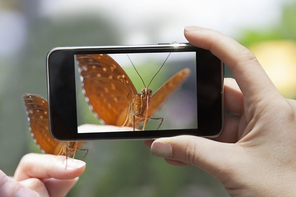
According to Chao, the technique and approach have been tested and shown to handle challenging recognition tasks, such as butterfly mimicries, in which species are differentiated by fine details and variety in their wing patterns and coloring. The ease with which the algorithm can be used could also allow imageomics to be integrated into a variety of other diverse purposes, ranging from climate to material science research, he said.

Chao said that one of the most challenging parts of fostering imageomics research is integrating different parts of scientific culture to collect enough data and form novel scientific hypotheses from them. That being said, he is enthusiastic about its potential to allow for the natural world to be seen within multiple fields.

“What we really want is for AI to have strong integration with scientific knowledge, and I would say imageomics is a great starting point towards that,” he said.


Bio Photonics Research Award


Visit: biophotonicsresearch.com
Nominate Now: https://biophotonicsresearch.com/award-nomination/?ecategory=Awards&rcategory=Awardee

#MeatAnalysis #FluorescenceTech #FoodQuality #FoodSafety #SpectroscopyInFood #MeatAuthentication #RapidDetection #FoodScience #MeatFreshness #MolecularDetection #FoodIndustryInnovation #NonDestructiveTesting #FoodMonitoring #SpectroscopyApplications #QualityControl #AdvancedSpectroscopy #MeatSpoilageDetection #FoodIntegrity #SmartFoodTesting #RealTimeAnalysis #FoodAuthenticity #FoodSafetyInnovation #SpectroscopyResearch #NextGenFoodSafety #InnovativeFoodScience,

Wednesday, July 9, 2025

Imaging Technology Shows How RNA in Cells Can Affect Health


Insight into the cellular distribution of RNA, which is closely linked to cell functions, could help scientists better understand the relation between cellular processes and disease. Potentially, this could lead to more targeted treatments for neurodegenerative disorders and aging.

While many methods have been developed to study RNA distribution within cells, only a few have been applied on a transcriptome-wide scale.

To capture the transcriptome of target cell types at the tissue level and RNA content within subcellular compartments, a research team at the UT Southwestern Medical Center, led by professor Haiqi Chen, developed Photoselection of Transcriptome over Nanoscale (PHOTON).

PHOTON combines high-resolution imaging with high-throughput sequencing to achieve spatial transcriptome profiling of RNA at subcellular resolution. It identifies RNA molecules at their native locations within cells, showing where different RNA species are distributed spatially in response to cellular cues.

To build PHOTON, the researchers designed DNA-based molecular cages that bound to all the RNA in cells. The molecular cages open when they are exposed to light, allowing for further chemical labeling.

After observing microscopically that the cells bound to the molecular cages, the researchers shined a narrow, 200-300-nm, near-ultraviolet (NUV) laser beam on regions of interest, such as specific organelles. The light caused the molecular cages to open, allowing only the RNA molecules located in the illuminated regions to be labeled. The researchers then collected the labeled RNA molecules and sequenced them to learn their identities and functions.

The team used PHOTON to examine RNAs present in the nucleolus and mitochondria, showing that RNAs identified through PHOTON closely matched those in published databases that were produced by isolating the organelles from the cells.

The researchers applied PHOTON to stress granules — transient, membraneless structures formed by cells when the cells are under stress. Although most stress granule RNAs that were identified matched those in published databases, the researchers found some discrepancies using PHOTON.

At the tissue scale, PHOTON accurately captured the transcriptome of cells within their native tissue microenvironment. At the subcellular scale, it enabled selective sequencing of the RNA content in the nucleoli, the mitochondria, and the stress granules.

The researchers used PHOTON to investigate whether m6A, a chemical modification found on some RNA molecules, played a role in moving RNAs into stress granules. By analyzing RNA molecules identified through PHOTON, the researchers found that the RNAs in the stress granules carried significantly more m6A than those outside the granules, suggesting that m6A contributes to the movement of specific RNAs into stress granules.

The researchers showed that PHOTON could be flexibly applied across regions of interest that spanned different scales, from specific regions of mouse ovarian tissue to various subcellular compartments. In-line image segmentation enabled the researchers to generate regions of interest based on an extensive range of spatial features, and automated the targeted photocleavage process over large numbers of cells or features.

These results show that PHOTON has the potential to uncover connections between spatial and transcriptomic information at diverse length scales.

Existing techniques to spatially identify RNA species can be prohibitively expensive and typically require specialized technical expertise and sophisticated image processing and data analysis to complete.

Chen said that he and his colleagues plan to use PHOTON to study the distributions of RNA in various conditions, particularly in neurodegenerative disease and aging. By comparing distributions in diseased cells to those in healthy cells, Chen said, researchers may be able to identify new targets for therapies to treat these conditions.

“Aging and many neurodegenerative diseases impose significant stress on cells, causing a subset of cellular RNA to redistribute into various subcellular compartments such as the stress granules,” Chen said. “PHOTON allows us to detect the spatial redistribution of cellular RNA in diseases versus health, helping us understand how these diseases cause damage to cellular functions.”

Bio Photonics Research Award

Visit: biophotonicsresearch.com
Nominate Now: https://biophotonicsresearch.com/award-nomination/?ecategory=Awards&rcategory=Awardee

#MeatAnalysis #FluorescenceTech #FoodQuality #FoodSafety #SpectroscopyInFood #MeatAuthentication #RapidDetection #FoodScience #MeatFreshness #MolecularDetection #FoodIndustryInnovation #NonDestructiveTesting #FoodMonitoring #SpectroscopyApplications #QualityControl #AdvancedSpectroscopy #MeatSpoilageDetection #FoodIntegrity #SmartFoodTesting #RealTimeAnalysis #FoodAuthenticity #FoodSafetyInnovation #SpectroscopyResearch #NextGenFoodSafety #InnovativeFoodScience,

24th Edition of World Biophotonics Research Awards 2026 | International Scientific Awards in Kuala Lumpur, Malaysia

  24th World Biophotonics Research Awards 2026: A Global Platform for Scientific Excellence and Innovation Science has always been the drivi...