Thursday, July 31, 2025

Spectrometer Measures from UV to NIR, Small Enough for Smartphone


Researchers at North Carolina State University (NCSU) have successfully demonstrated a spectrometer that is orders of magnitude smaller than current technologies and can accurately measure wavelengths of light from ultraviolet to the near-infrared (NIR). The technology makes it possible to create hand-held spectroscopy devices and holds promise for the development of devices that incorporate an array of the new sensors to serve as next-generation imaging spectrometers, the researchers said.

Spectrometers are used in applications ranging from manufacturing to biomedical diagnostics to discern the chemical and physical properties of various materials based on how light changes as it interacts with those materials. Despite their widespread use, even the smallest spectrometers on the market are fairly cumbersome.

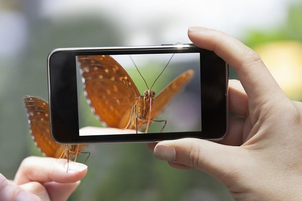
“We’ve created a spectrometer that operates quickly, at low voltage, and that is sensitive to a wide spectrum of light,” said Brendan O’Connor, corresponding author of a paper on the work and a professor of mechanical and aerospace engineering at NCSU. “Our demonstration prototype is only a few square millimeters in size — it could fit on your phone. You could make it as small as a pixel, if you wanted to.”

The technology makes use of a tiny photodetector capable of sensing wavelengths of light after the light interacts with a target material. Applying different voltages to the photodetector manipulates which wavelengths of light it is most sensitive to.

“If you rapidly apply a range of voltages to the photodetector, and measure all of the wavelengths of light being captured at each voltage, you have enough data that a simple computational program can recreate an accurate signature of the light that is passing through or reflecting off of the target material,” O’Connor said. “The range of voltages is less than one volt, and the entire process can take place in less than a millisecond.”

Previous attempts to create miniaturized photodetectors have relied on complex optics, used high voltages, or have not been as sensitive to such a broad range of wavelengths.

In proof-of-concept testing, the researchers found their pixel-sized spectrometer was as accurate as a conventional spectrometer and had sensitivity comparable to commercial photodetection devices.

“In the long term, our goal is to bring spectrometers to the consumer market,” O’Connor said. “The size and energy demand of the technology make it feasible to incorporate into a smartphone, and we think this makes some exciting applications possible. From a research standpoint, this also paves the way for improved access to imaging spectroscopy, microscopic spectroscopy, and other applications that would be useful in the lab.

Bio Photonics Research Award

Visit: biophotonicsresearch.com
Nominate Now: https://biophotonicsresearch.com/award-nomination/?ecategory=Awards&rcategory=Awardee

#MeatAnalysis #FluorescenceTech #FoodQuality #FoodSafety #SpectroscopyInFood #MeatAuthentication #RapidDetection #FoodScience #MeatFreshness #MolecularDetection #FoodIndustryInnovation #NonDestructiveTesting #FoodMonitoring #SpectroscopyApplications #QualityControl #AdvancedSpectroscopy #MeatSpoilageDetection #FoodIntegrity #SmartFoodTesting #RealTimeAnalysis #FoodAuthenticity #FoodSafetyInnovation #SpectroscopyResearch #NextGenFoodSafety #InnovativeFoodScience,

Wednesday, July 30, 2025

Lasers Improve Phototherapy-Drug Delivery Platform


Photoimmunotherapy targets cancer cells with microscopic, nano-engineered cancer drugs that are light-activated at the lesion site. Although the technology is increasingly used to treat metastatic cancer, tools to improve the effectiveness of photoimmunotherapy in gynecologic oncology have been lacking. Responses to treatment vary from person to person, and no method has existed to readily monitor whether the drugs are delivered effectively or have the desired therapeutic effect.

Researchers from the University of Maryland (UMD), in collaboration with medical laser manufacturer Modulight, demonstrated that the efficacy, safety, and consistency of photoimmunotherapy can be improved by integrating targeted, light-based techniques for drug delivery with laser-assisted endoscopy and fluorescence-guided treatment planning.

The researchers designed a drug delivery system using targeted, photo-activable, multi-agent liposomes (TPMAL). TPMAL consists of chemotherapy drugs that are labeled with fluorescent markers. It improves therapeutic delivery and monitors the growth of metastases in difficult-to-treat locations within the body.

A laser-assisted endoscopic camera created by Modulight captures and uploads information from TPMAL to Modulight Cloud, a cloud services platform. Modulight Cloud analyzes the multispectral fluorescence emission from TPMAL in real time to enable fluorescence-guided drug delivery and fluorescence-guided light dosimetry. The real-time connectivity enables simultaneous treatment monitoring, remote protocol development, and rapid transfer and viewing of data.

The data acquired by the camera can be used to customize fluorescence-guided therapy. Doctors can use the data to determine, in real time, how well the drug-carrying liposomes are reaching and delivering treatment to metastatic cancer sites. They can also use the data to calculate the precise amount of laser light needed during photoimmunotherapy to minimize tissue damage.

The optimal accumulation of TPMAL in tumors can occur at different times for different patients. Fluorescence-guided drug delivery identifies the time when the TPMAL fluorescence signal is near peak levels in tumors, thereby informing the initiation of photoimmunotherapy for the best outcome.

The TPMAL levels in tumors can vary among patients. Fluorescence-guided light dosimetry monitors the photobleaching of photosensitizers during photoimmunotherapy, so that the light dose can be adjusted accordingly, in real time, to improve the consistency of treatment effects.

“Patients undergoing cancer treatment exhibit varying tolerances and individualized responses,” said UMD professor Huang Chiao Huang. “Through continuous monitoring of their progress, we can promptly determine if treatment adjustments are needed while they are actively receiving care. There is no need to postpone decisions until the next scheduled session.”

The researchers demonstrated their approach in mouse models with peritoneal carcinomatosis, a type of cancer that can be caused by metastatic ovarian cancer. They observed a fourteenfold improvement in drug delivery to metastatic tumors. The information captured by the endoscopic camera prompted fluorescence-guided light dosimetry in over 50% of the mice. By combining TPMAL, laser-assisted imaging, and fluorescence-guided intervention, the researchers achieved a more homogeneous response to treatment in the mice and improved tumor control without causing side effects.

Although nearly half of the women with advanced ovarian cancer achieve complete remission after surgery and chemotherapy, many patients will relapse due largely to residual submillimeter lesions. These residual micro-metastases are difficult to detect and often develop resistance to standard treatments. New approaches are needed to address these drug-resistant micro-metastases.

The UMD-Modulight study shows that a combination of targeted photomedicine drug delivery, imaging, and monitoring of treatment responses can help prevent the return and spread of cancer while minimizing the side effects of treatment. Together, the three techniques can give doctors the ability to make immediate adjustments in treatment to achieve better outcomes.

“This approach ensures that the treatment remains both effective and uniform,” Huang said. “This process is repeated as necessary for each patient, resulting in a personalized dosage tailored to maximize treatment effectiveness within a single session.”

The team is now working to establish a comprehensive panel of biomarkers that would allow doctors to promptly determine the optimal dosage for each patient during a treatment session.

“The goal is to minimize the number of treatment cycles required while minimizing burden on the patient’s body, in contrast to conventional treatment approaches,” Huang said.

Bio Photonics Research Award


Visit: biophotonicsresearch.com
Nominate Now: https://biophotonicsresearch.com/award-nomination/?ecategory=Awards&rcategory=Awardee

#MeatAnalysis #FluorescenceTech #FoodQuality #FoodSafety #SpectroscopyInFood #MeatAuthentication #RapidDetection #FoodScience #MeatFreshness #MolecularDetection #FoodIndustryInnovation #NonDestructiveTesting #FoodMonitoring #SpectroscopyApplications #QualityControl #AdvancedSpectroscopy #MeatSpoilageDetection #FoodIntegrity #SmartFoodTesting #RealTimeAnalysis #FoodAuthenticity #FoodSafetyInnovation #SpectroscopyResearch #NextGenFoodSafety #InnovativeFoodScience,

Tuesday, July 29, 2025

Holographic Imaging Measures Cellular Structures without Distorting Them


Biomolecular condensates — membraneless, microscopic structures that concentrate proteins and other molecules in cells — are crucial to the organization of cellular biochemistry. Insight into the development and behavior of condensates could lead to better treatments for infectious diseases, cancer, and neurological disorders.

Researchers at New York University (NYU) aimed to measure condensate composition and dynamics without relying on conventional techniques, like fluorescence labeling or surface attachment, which can damage fragile condensate samples. Until now, scientists have needed to distort condensate samples to study them.

“It’s been the elephant in the room for scientists,” professor Saumya Saurabh said. “Our research provides a precise and noninvasive way to study biomolecular condensates.”

To overcome the limitations of conventional techniques, the team used label-free holographic microscopy to investigate the behavior of a condensate-forming protein in vitro. The researchers flowed thousands of droplets through a holographic microscope in a microfluidic channel to visualize and characterize each particle individually. The holographic characterization was free from perturbations and was able to gather data on thousands of particles in minutes. Precise information about the droplet’s size, shape, and refractive index was encoded in the hologram of each μm-scale droplet.

The researchers used this technique to examine PopZ, a condensate-forming protein that influences cell growth. The precision and speed provided by the digital holography technique enabled the team to monitor the kinetics of the condensate’s formation, growth, and aging over time.

By systematically varying the concentration and valence of cations, the researchers found that multivalent ions influence condensate organization and dynamics. “I was surprised by their complex and incredibly sensitive response to different ionic species,” researcher Julian von Hofe said. “Even a small change in ionic valency drastically altered both condensate concentration and dynamics.”

The researchers used superresolution microscopy to explore the architecture of PopZ at the nanoscale. Data acquired through superresolution imaging revealed that the condensates were not uniform droplets, but exhibited intricate nanoscale organization, and that PopZ droplet growth deviated from classical models. These findings were supported by molecular dynamics simulations, which provided atomic-level insights into the biocondensate assemblies.

The study thus demonstrated the value of holographic microscopy as a hypothesis-generating tool that provides noninvasive insight into condensate substructure, that can be further tested and refined using complementary, minimally perturbative methods.

“Being able to see ‘under the hood’ for the first time has revealed some big surprises about this important class of systems,” professor David Grier said.

Although the researchers observed the condensates in vitro, their findings could contribute to a more complete understanding of condensate behavior within living cells. “The intricate reality of biomolecular condensates, as revealed by our findings, goes far beyond simple liquid-liquid phase separation,” Saurabh said.

A better understanding of how biomolecular condensates are organized and grow, made possible through holographic microscopy and superresolution imaging, could help shape disease modeling and future drug development. For example, the proteins that form plaques in ALS are fluid condensates in good health. “Understanding how a spherical condensate forms into a deadly plaque is an opportunity to better understand ALS,” Saurabh said.

The biomolecular condensates in the cells can also house drug molecules that are intended for a different purpose. This phenomenon could help explain why drugs that are designed to target a specific protein still cause side effects. By using holographic microscopy to analyze condensate dynamics with extreme precision, scientists can identify the subtle differences in condensate composition and architecture that occur when drug molecules enter a condensate.

“For example, we can now explore the chemical space of drug modifications to precisely control their partitioning, achieving the specificity needed to prevent them from entering condensates,” Saurabh said. “This opens new avenues for how we think about designing drugs and their potential side effects.”

This work highlights the power of holographic microscopy, especially when used with superresolution imaging, to probe the properties and mechanistic underpinnings of biomolecular condensates. “Our collaboration has introduced fast, precise, and effective methods for measuring the composition and dynamics of macromolecular condensates,” Grier said.


Bio Photonics Research Award


Visit: biophotonicsresearch.com
Nominate Now: https://biophotonicsresearch.com/award-nomination/?ecategory=Awards&rcategory=Awardee

#MeatAnalysis #FluorescenceTech #FoodQuality #FoodSafety #SpectroscopyInFood #MeatAuthentication #RapidDetection #FoodScience #MeatFreshness #MolecularDetection #FoodIndustryInnovation #NonDestructiveTesting #FoodMonitoring #SpectroscopyApplications #QualityControl #AdvancedSpectroscopy #MeatSpoilageDetection #FoodIntegrity #SmartFoodTesting #RealTimeAnalysis #FoodAuthenticity #FoodSafetyInnovation #SpectroscopyResearch #NextGenFoodSafety #InnovativeFoodScience,

Monday, July 28, 2025

Handheld Device Allows Imaging and Treatment of Oral Cancer


Oral cancer is a growing public health concern, particularly in South Asia, where it affects tens of thousands each year. In India alone, oral cancer accounts for 40% of all cancers, largely driven by the widespread use of tobacco-based products. The situation is worsened by limited access to early screening and treatment, especially in rural and underserved areas. Most cases are diagnosed at advanced stages, when treatment is more difficult and survival rates are lower.

To address this problem, a team of researchers has developed a compact, affordable device that can both image suspicious lesions and deliver light-based therapy to treat them.

The device uses a smartphone-coupled intraoral probe with specialized LEDs and filters to capture white-light and fluorescence images to pinpoint oral cancers. It also includes laser diodes to activate a light-sensitive compound called protoporphyrin IX (PpIX), which accumulates in cancerous tissue after the application of a precursor drug, 5-aminolevulinic acid (ALA). When exposed to light, PpIX produces reactive molecules that destroy cancer cells while sparing healthy tissue. This approach, known as photodynamic therapy, has shown promise in treating early oral cancers with minimal side effects.

To evaluate the device, the researchers conducted a series of preclinical tests. The team used tissue-mimicking phantoms and cell cultures to test the device’s ability to detect PpIX fluorescence and monitor its breakdown (photobleaching) during treatment. The device showed a strong linear response to increasing PpIX concentrations and could detect changes in fluorescence that corresponded to effective light dosing.

Simulated 3D oral tissues embedded with cancer cells were used to assess how deeply the device could detect and treat lesions. The system successfully imaged PpIX fluorescence up to 2.5 mm deep and showed effective photobleaching at depths relevant to early-stage oral cancers. To further test the technology, the device was used to deliver photodynamic therapy and monitor treatment in an animal model. There, tumors treated with the device shrank significantly compared to untreated controls. Histological analysis revealed tumor cell death extending up to 3.5 mm deep, consistent with light delivery simulations.

One of the device’s key features is its capability to monitor treatment in real time. By measuring the decrease in PpIX fluorescence during light exposure, the system provides feedback on how much therapeutic dose has been delivered. This could help ensure that each treatment is effective, even in settings without advanced medical infrastructure.

The researchers also used ratiometric imaging — comparing red and green fluorescence signals — to improve the accuracy of lesion detection and treatment monitoring. This method helps distinguish cancerous tissue from surrounding healthy areas, even in complex tissue environments.

The study demonstrates that a low-cost, portable device can perform both diagnosis and treatment of early oral cancer with promising accuracy and effectiveness. By combining imaging and therapy in a single tool, the technology could streamline care in regions where access to specialists is limited.

Future work will focus on clinical trials and refining the device for broader use. The team envisions a system that not only guides treatment but also adapts in real time, making photodynamic therapy more accessible and effective for patients around the world.


Bio Photonics Research Award

Visit: biophotonicsresearch.com
Nominate Now: https://biophotonicsresearch.com/award-nomination/?ecategory=Awards&rcategory=Awardee

#MeatAnalysis #FluorescenceTech #FoodQuality #FoodSafety #SpectroscopyInFood #MeatAuthentication #RapidDetection #FoodScience #MeatFreshness #MolecularDetection #FoodIndustryInnovation #NonDestructiveTesting #FoodMonitoring #SpectroscopyApplications #QualityControl #AdvancedSpectroscopy #MeatSpoilageDetection #FoodIntegrity #SmartFoodTesting #RealTimeAnalysis #FoodAuthenticity #FoodSafetyInnovation #SpectroscopyResearch #NextGenFoodSafety #InnovativeFoodScience,

Saturday, July 26, 2025

Material Innovation Keys Scalable Spectrometer Design for Diverse Applications


A smartphone-inspired spectrometer platform, built with low-cost plastic materials instead of glass, could make spectral imaging more accessible across the scientific, industrial, and consumer domains.

The spectrometer spans the visible to SWIR range and is fabricated using mass-producible, non-lithographic methods. These properties could make it suitable for in-home health care monitoring, food quality testing, agricultural sensing, and many other applications that require affordable, broadband sensing capabilities.

The spectrometer design is the result of a collaboration among researchers at the University of Cambridge, Zhejiang University, Zhejiang Sci-Tech University, and Nanyang Technological University, with backgrounds in materials science, optical engineering, and signal processing.

Plastic optical components are used in smartphone cameras to achieve high performance in an ultracompact format. Inspired by this approach, the research team took a similar path, using transparent shape memory epoxies to stress-engineer optical dispersive elements made from plastic. The epoxy used for the spectrometer, bisphenol A epoxy, is highly transparent across the visible to SWIR range.

Shape memory epoxies can be mechanically stretched at elevated temperatures to program precise, stable stress distributions into the material. These stresses create birefringence, an optical effect where light is split according to its wavelength.

Through temperature-controlled mechanical stretching, the team was able to stress-engineer the epoxy and tailor its optical properties. Shape memory epoxies provide superior stress storage compared to other plastic materials, which enables a wide range of spectral encoding through stress engineering.

“By shaping the internal stress within the polymer, we are able to engineer spectral behavior with high repeatability and tunability, something that’s incredibly difficult to achieve with conventional optics,” professor Gongyuan Zhang said.

The resulting films act as spectral filters, encoding information that can be read by standard CMOS image sensors and reconstructed via algorithms. The researchers demonstrated that the planar, stress-engineered epoxy films can be used to form a spectrometer device when they are integrated with a commercial CMOS image sensor and a spectral reconstruction algorithm is used for computational processing of the pixel outputs.

The use of large-scale stretched epoxy films as filters significantly enhances the yield of the spectrometer. The team realized miniaturized spectrometers with broad coverage across both the visible (400-800 nm) and NIR (800-1600 nm) ranges.

The epoxy film layer also enables the spectrometer to serve as a line-scanning device for spectral imaging on 2D images, facilitating the acquisition of corresponding spectral data cubes, and demonstrating the spectrometer’s potential as a portable tool for hyperspectral imaging.

The stress-engineered films can be fabricated in a single step, without the need for lithography or expensive nanofabrication, making the spectrometers suitable for mass production and integration into consumer electronics like mobile phones and wearable technologies.

“We’ve shown that you can use programmable plastics to cover a much broader range of the spectrum than typical miniaturized systems — right into the SWIR,” professor Zongyin Yang said. “That’s really important for applications like agricultural monitoring, mineral exploration, and medical diagnostics.”

The new spectrometer design could be used to detect pollutants, verify the authenticity of drugs, monitor blood sugar noninvasively, and even to sort recyclable materials in real-time. By eliminating the trade-offs between size, cost, and spectral range, the spectrometer could help advance research in computational photonics and sustainable sensing technologies.

“This work shows how mechanical design principles can be used to reshape photonic functionality,” professor Tawfique Hasan said. “By embedding stress into transparent polymers, we have created a new class of dispersive optics that are not only lightweight and scalable but also adaptable across a wide spectral range. This level of flexibility is very difficult to achieve with traditional optics relying on static, lithographically defined structures.”

As the team continues to refine the design and explore commercial pathways, the stress-engineered, plastic spectrometer could become a building block for the next generation of intelligent, compact sensors embedded in devices for everyday use.


Bio Photonics Research Award

Visit: biophotonicsresearch.com
Nominate Now: https://biophotonicsresearch.com/award-nomination/?ecategory=Awards&rcategory=Awardee

#MeatAnalysis #FluorescenceTech #FoodQuality #FoodSafety #SpectroscopyInFood #MeatAuthentication #RapidDetection #FoodScience #MeatFreshness #MolecularDetection #FoodIndustryInnovation #NonDestructiveTesting #FoodMonitoring #SpectroscopyApplications #QualityControl #AdvancedSpectroscopy #MeatSpoilageDetection #FoodIntegrity #SmartFoodTesting #RealTimeAnalysis #FoodAuthenticity #FoodSafetyInnovation #SpectroscopyResearch #NextGenFoodSafety #InnovativeFoodScience,


Thursday, July 24, 2025

Researchers Shrink Titanium-Sapphire Laser to Chip Scale


Lasers based on titanium-sapphire (Ti:sapphire) provide top performance in fields like quantum optics, spectroscopy, and neuroscience. But that performance comes at a steep cost of not just the multi-thousand dollar price tag, but space and power as well. Ti:sapphire lasers take up several cubic feet and require other high-powered lasers to supply them with enough energy to function. Despite their high level of performance and utility in cutting edge applications, their adoption in the industry has been slow.

Making a jump from tabletop to the microscale, engineers at Stanford University have built a Ti:sapphire laser on a chip. According to the researchers, the prototype is four orders of magnitude smaller (10,000×) and three orders less expensive (1,000×) than any Ti:sapphire laser ever produced.

“Instead of one large and expensive laser, any lab might soon have hundreds of these valuable lasers on a single chip. And you can fuel it all with a green laser pointer,” said Jelena Vuckovic, Stanford’s Jensen Huang Professor in Global Leadership and senior author of the research.

To fashion the new laser, the researchers began with a bulk layer of Ti:sapphire on a platform of silicon dioxide (SiO2), all riding atop true sapphire crystal. They then grind, etch, and polish the Ti:sapphire to an extremely thin layer just a few hundred nanometers thick. Into that thin layer, they then pattern a swirling vortex of tiny ridges, or a waveguide. These ridges are like fiber-optic cables, guiding the light around and around, building in intensity.

“Mathematically speaking, intensity is power divided by area. So, if you maintain the same power as the large-scale laser, but reduce the area in which it is concentrated, the intensity goes through the roof,” said Joshua Yang, a doctoral candidate in Vuckovic’s lab and co-first author. “The small scale of our laser actually helps us make it more efficient.”

A microscale heater that warms the light traveling through the waveguides is then added, allowing the team to change the wavelength of the emitted light to tune the color of the light anywhere between 700 nm and 1,000 nm – in the red to infrared range.

“When you leap from tabletop size and make something producible on a chip at such a low cost, it puts these powerful lasers in reach for a lot of different important applications,” Yang said.

These applications include areas like quantum physics, where the laser could provide an inexpensive and practical solution to scale down state-of-the-art quantum computers. Medical fields could see the Ti:sapphire lasers being used for optogenetics or in compact optical coherence tomography technologies for ophthalmology.

The researchers are currently working on next steps for perfecting their chip-scale Ti:sapphire laser as well as on ways to mass-produce them on wafers and bring them to market.

Bio Photonics Research Award


Visit: biophotonicsresearch.com
Nominate Now: https://biophotonicsresearch.com/award-nomination/?ecategory=Awards&rcategory=Awardee

#MeatAnalysis #FluorescenceTech #FoodQuality #FoodSafety #SpectroscopyInFood #MeatAuthentication #RapidDetection #FoodScience #MeatFreshness #MolecularDetection #FoodIndustryInnovation #NonDestructiveTesting #FoodMonitoring #SpectroscopyApplications #QualityControl #AdvancedSpectroscopy #MeatSpoilageDetection #FoodIntegrity #SmartFoodTesting #RealTimeAnalysis #FoodAuthenticity #FoodSafetyInnovation #SpectroscopyResearch #NextGenFoodSafety #InnovativeFoodScience,

Wednesday, July 23, 2025

Handheld Device Allows Imaging and Treatment of Oral Cancer


Oral cancer is a growing public health concern, particularly in South Asia, where it affects tens of thousands each year. In India alone, oral cancer accounts for 40% of all cancers, largely driven by the widespread use of tobacco-based products. The situation is worsened by limited access to early screening and treatment, especially in rural and underserved areas. Most cases are diagnosed at advanced stages, when treatment is more difficult and survival rates are lower.

To address this problem, a team of researchers has developed a compact, affordable device that can both image suspicious lesions and deliver light-based therapy to treat them.

The device uses a smartphone-coupled intraoral probe with specialized LEDs and filters to capture white-light and fluorescence images to pinpoint oral cancers. It also includes laser diodes to activate a light-sensitive compound called protoporphyrin IX (PpIX), which accumulates in cancerous tissue after the application of a precursor drug, 5-aminolevulinic acid (ALA). When exposed to light, PpIX produces reactive molecules that destroy cancer cells while sparing healthy tissue. This approach, known as photodynamic therapy, has shown promise in treating early oral cancers with minimal side effects.

Simulated 3D oral tissues embedded with cancer cells were used to assess how deeply the device could detect and treat lesions. The system successfully imaged PpIX fluorescence up to 2.5 mm deep and showed effective photobleaching at depths relevant to early-stage oral cancers. To further test the technology, the device was used to deliver photodynamic therapy and monitor treatment in an animal model. There, tumors treated with the device shrank significantly compared to untreated controls. Histological analysis revealed tumor cell death extending up to 3.5 mm deep, consistent with light delivery simulations.

One of the device’s key features is its capability to monitor treatment in real time. By measuring the decrease in PpIX fluorescence during light exposure, the system provides feedback on how much therapeutic dose has been delivered. This could help ensure that each treatment is effective, even in settings without advanced medical infrastructure.

The researchers also used ratiometric imaging — comparing red and green fluorescence signals — to improve the accuracy of lesion detection and treatment monitoring. This method helps distinguish cancerous tissue from surrounding healthy areas, even in complex tissue environments.

The study demonstrates that a low-cost, portable device can perform both diagnosis and treatment of early oral cancer with promising accuracy and effectiveness. By combining imaging and therapy in a single tool, the technology could streamline care in regions where access to specialists is limited.

Future work will focus on clinical trials and refining the device for broader use. The team envisions a system that not only guides treatment but also adapts in real time, making photodynamic therapy more accessible and effective for patients around the world.


Bio Photonics Research Award

Visit: biophotonicsresearch.com
Nominate Now: https://biophotonicsresearch.com/award-nomination/?ecategory=Awards&rcategory=Awardee

#MeatAnalysis #FluorescenceTech #FoodQuality #FoodSafety #SpectroscopyInFood #MeatAuthentication #RapidDetection #FoodScience #MeatFreshness #MolecularDetection #FoodIndustryInnovation #NonDestructiveTesting #FoodMonitoring #SpectroscopyApplications #QualityControl #AdvancedSpectroscopy #MeatSpoilageDetection #FoodIntegrity #SmartFoodTesting #RealTimeAnalysis #FoodAuthenticity #FoodSafetyInnovation #SpectroscopyResearch #NextGenFoodSafety #InnovativeFoodScience,

Tuesday, July 22, 2025

Ultrafast Microscope Images Material’s Molecular Structure and Dynamics


Perovskites, a family of organic-inorganic hybrid materials, are efficient at converting light to electricity and relatively easy to make. They absorb certain colors of the visible spectrum effectively and can be layered with other materials, such as silicon, that absorb wavelengths the perovskites cannot capture.

But the low photostability of perovskites makes it difficult to improve their performance. Perovskite solar cells can be made from many different combinations of chemical compositions and prepared under various conditions. It is hard to predict how these factors will affect the structure and performance of the perovskite cell.

Many complex materials found in semiconductors, displays, and quantum and biomedical applications present the same challenge. To better understand how to improve these materials, scientists need to be able to visualize the material’s dynamics at the subatomic, atomic, and molecular levels.

A research team at the University of Colorado-Boulder (CU Boulder) developed a microscope for spatio-spectral-temporal ultrafast nanoimaging of the structural characteristics of materials. The ultrafast microscope enables researchers to directly image the role of molecular order, disorder, and local crystallinity in the optical and electronic properties of materials.

The researchers used the microscope to perform combined ground- and ultrafast excited-state IR nanoimaging of a metal halide perovskite that is a promising candidate for tandem solar cells, photocatalysis, and optoelectronic applications.

The microscope is equipped with a metal-coated nanotip that is positioned within a nanometer of the perovskite layer, then hit with a sequence of ultrashort laser pulses. The first pulse excites the electrons in the material in the visible, and subsequent pulses in the IR capture the movement and interaction of the electrons and molecules in the material over time. The nanotip functions like an antenna for the laser light, focusing the laser to the nanoscale.

The researchers scanned the nanotip across the perovskite layer, creating an image of the material pixel by pixel. Each image was the equivalent of one movie frame, due to the temporal differences in the laser pulses.

To reduce noise, the researchers used optical amplification techniques and developed a method to modulate the laser beams. “If you shine a light on this very tiny tip, the light that comes back is very weak since it only interacts with very few electrons or molecules,” researcher Branden Esses said. “It’s so weak that you need special techniques to detect it.”

According to researcher Roland Wilcken, controlling the way the light is focused at the nanometer scale and how it is emitted and detected is essential to achieving the contrast and signal necessary to make an ultrafast movie of the material.

The researchers captured ultrahigh-resolution images of atomic and molecular movement in the perovskite at the femtosecond scale and measured atomic motion in the molecules with very high precision. The photoexcited electrons and coupled changes of the lattice structure (i.e., polarons) were diagnosed spectroscopically with ultrahigh spatiotemporal resolution, enabling the researchers to better understand the perovskite’s structure and composition and its performance as a photovoltaic material.

The team’s findings suggest that the more disorder in the material, the better the photovoltaic performance. “In contrast to conventional semiconductors, it seems that more structural disorder gives rise to more stable photogenerated electrons in hybrid perovskites,” professor Markus Raschke said.

According to Raschke, there is limited knowledge of the processes that occur after sunlight is absorbed by photovoltaic materials, and how the excited electrons move in the material without being dispersed.

“We like to say that we’re making ultrafast movies,” he said. “For the first time, we can actually sort this out, because we can record spatial, temporal, and spectral dimensions simultaneously in this microscope.”

The team expects the ultrafast microscope to have a significant impact on the ability of material scientists to improve the performance of new semiconductor and quantum materials for computing, energy, and medical applications.

“This is a way to examine the material properties on a very elementary level, so that in the future we’ll be able to design materials with certain properties in a more directed way,” professor Sean Shaheen said.

“We’re able to say, ‘We know we prefer this kind of structure, which results in, for example, longer-lived electronic excitations as linked to photovoltaic performance,’ and then we’re able to inform our material synthesis partners to help make them,” Esses said.

Bio Photonics Research Award

Visit: biophotonicsresearch.com
Nominate Now: https://biophotonicsresearch.com/award-nomination/?ecategory=Awards&rcategory=Awardee

#MeatAnalysis #FluorescenceTech #FoodQuality #FoodSafety #SpectroscopyInFood #MeatAuthentication #RapidDetection #FoodScience #MeatFreshness #MolecularDetection #FoodIndustryInnovation #NonDestructiveTesting #FoodMonitoring #SpectroscopyApplications #QualityControl #AdvancedSpectroscopy #MeatSpoilageDetection #FoodIntegrity #SmartFoodTesting #RealTimeAnalysis #FoodAuthenticity #FoodSafetyInnovation #SpectroscopyResearch #NextGenFoodSafety #InnovativeFoodScience,

Monday, July 21, 2025

Photodynamic Method Boosts Antibiotics Against Drug-Resistant Bacteria


Multidrug-resistant bacteria are considered a serious threat to infection control. Faced with the increasing difficulty of developing new antibiotics to combat resilient bacterial strains, scientists are turning to photodynamic inactivation (PDI), a light-based approach to breaking antimicrobial resistance.

PDI strengthens the effect of antibiotics and induces oxidative stress in microorganisms through the interaction of light with a photosensitizer. The photosensitizer is energized in PDI by absorbing visible light to form reactive oxygen species that trigger bacterial inactivation by oxidizing and destroying microorganisms or weakening their resistance to antibiotics.

In recent work, researchers at the University of São Paulo’s Optics and Photonics Research Center showed that PDI can modify bacterial sensitivity to antibiotics and reduce the resistance and persistence of both standard and clinical strains. The researchers, led by professor Vanderlei Salvador Bagnato, investigated the effects of photodynamic action on resistant bacteria collected from patients and bacterial cells with laboratory-induced resistance. They focused their investigation on Staphylococcus aureus, a bacterium that causes a range of diseases, from skin infections to pneumonia.

The researchers used 10 μM, 10 J/cm2 of the photosensitizer curcumin at 450 nm with antibiotics. Curcumin has been shown to strengthen some antibiotics by affecting the bacterial membrane and other cellular components. Three antibiotics — amoxicillin, erythromycin, and gentamicin — were treated with curcumin.

The results showed that five cycles of PDI were sufficient to break bacterial resistance. The researchers found that S. aureus was most susceptible to gentamicin, although the other two antibiotics also proved effective against the bacteria after treatment with PDI.

In addition, the researchers concluded that a reduction in the degree of antimicrobial resistance through photo-oxidative action can mitigate antibiotic failures. Photodynamic action not only improves infection control, but also modifies the degree to which antimicrobials are susceptible to bacteria by decreasing bacterial resistance to below breakpoints and controlling the biofilm formation, which is an important bacterial virulence factor.

“We discovered that PDI doesn’t always destroy the bacteria, but it does destroy part of the mechanisms they use to become drug-resistant,” Bagnato said. “This led to the idea of trying an oxidative shock to make them susceptible to antibiotics.”

Historically, the primary strategy in the fight against multidrug-resistant bacteria infections has been the development of new antimicrobial drugs. However, failures in antibiotic treatments occur. Antibiotics act on a certain bacterial cell compartment, depending on their type, while PDI acts in the entire bacterial cell, causing multiple damages.

The researchers said that an increase or a complete recovery of a bacterium’s susceptibility to antibiotics could be a way to prolong the useful life of recent classes of antibiotics, which is essential to avoid infection control collapses. They said that a broader assessment of other antibiotics, microorganisms, and photosensitizers is needed to achieve a full understanding of the mechanisms of action and interaction between PDI and antibiotics.

Bio Photonics Research Award

Visit: biophotonicsresearch.com
Nominate Now: https://biophotonicsresearch.com/award-nomination/?ecategory=Awards&rcategory=Awardee

#MeatAnalysis #FluorescenceTech #FoodQuality #FoodSafety #SpectroscopyInFood #MeatAuthentication #RapidDetection #FoodScience #MeatFreshness #MolecularDetection #FoodIndustryInnovation #NonDestructiveTesting #FoodMonitoring #SpectroscopyApplications #QualityControl #AdvancedSpectroscopy #MeatSpoilageDetection #FoodIntegrity #SmartFoodTesting #RealTimeAnalysis #FoodAuthenticity #FoodSafetyInnovation #SpectroscopyResearch #NextGenFoodSafety #InnovativeFoodScience,

Saturday, July 19, 2025

Squishy Lasers Could Reveal Secrets of Cell Growth Origins


Researchers at the University of St. Andrews and the University of Cologne have developed lasers that they have described as “squishy.” These devices could help solve the biological mysteries behind the development of embryos and cancerous tumors.

Fundamental biological processes driven by mechanical forces invisible to the naked eye are currently poorly understood by scientists. The squishy lasers developed by the researchers are able to precisely measure the forces exerted by biological cells.

“Embryos and tumors both start with just a few cells,” said professor Malte Gather from the University of St. Andrews. “It is still very challenging to understand how they expand, contract, squeeze, and fold as they develop. Being able to measure biological forces in real-time could be transformative. It could hold the key to understanding the exact mechanics behind how embryos develop, whether successfully or unsuccessfully, and how cancer grows.”

These squishy microlasers can be injected directly into embryos or mixed into artificial tumors. According to Marcel Schubert, a professor at the University of Cologne, the microlasers are actually droplets of oil doped with fluorescent dye.

“As the biological forces get to work, the microlasers are squished and deformed by the cells around them. The laser light changes its color in response and reveals the force that’s acting upon it,” Schubert said.

The innovation allows researchers to measure and monitor biological forces in real time, Schubert said. Additionally, he said, it works in thick biological tissue, an area where other methods would require an almost transparent sample.

The oil and fluorescent dye used to create the microlasers are made from nontoxic, readily available materials, ensuring they do not interfere with biological processes. This aspect makes the technology not only effective but also commercially viable.

The researchers tested their method on fruit fly larvae, to see how they developed, as well as in artificial tumors made from brain tumor cells, so-called tumor spheroids.

“We measured the 3D distribution of forces within tumor spheroids and made high-resolution long-term force measurements within the fruit fly larvae,” Gather said.

The team is now seeking funding to adapt their method for clinical trials, aiming to extend its application to larger cell systems.

Bio Photonics Research Award

Visit: biophotonicsresearch.com
Nominate Now: https://biophotonicsresearch.com/award-nomination/?ecategory=Awards&rcategory=Awardee

#MeatAnalysis #FluorescenceTech #FoodQuality #FoodSafety #SpectroscopyInFood #MeatAuthentication #RapidDetection #FoodScience #MeatFreshness #MolecularDetection #FoodIndustryInnovation #NonDestructiveTesting #FoodMonitoring #SpectroscopyApplications #QualityControl #AdvancedSpectroscopy #MeatSpoilageDetection #FoodIntegrity #SmartFoodTesting #RealTimeAnalysis #FoodAuthenticity #FoodSafetyInnovation #SpectroscopyResearch #NextGenFoodSafety #InnovativeFoodScience,

Friday, July 18, 2025

Raman Approach Safely Tracks Live-Cell RNA Expression


Single-cell RNA sequencing enables scientists to interrogate cells at extraordinary resolution and scale. However, the sequencing process destroys the cell, making it difficult to use the technique to study ongoing changes in gene expression.

Raman microscopy measures the vibrational energy levels of proteins and metabolites in a nondestructive manner at subcellular spatial resolution, but it is unable to interpret genetic information.

“RNA sequencing gives you extremely detailed information, but it’s destructive,” researcher Koseki Kobayashi-Kirschvink said. “Raman is noninvasive, but it doesn’t tell you anything about RNA.”

A new technique developed at MIT combines the advantages of single-cell RNA sequencing and Raman spectroscopy to track a cell’s RNA expression without damaging the cell. Known as Raman2RNA (R2R), this technique could allow scientists to study long-term cellular processes, such as cancer progression and embryonic development, using the same cells repeatedly.

To create the technique, the team trained a computational model to translate Raman signals into RNA expression states. “The idea of this project was to use machine learning to combine the strength of both modalities, thereby allowing you to understand the dynamics of gene expression profiles at the single-cell level over time,” Kobayashi-Kirschvink said.

To generate the data needed to train the machine learning model, the researchers treated mouse fibroblast cells with factors that reprogrammed the cells, causing them to become pluripotent (i.e., undifferentiated) stem cells. Using Raman spectroscopy, the team imaged the cells at 36 time points, over an 18-day period. During that time, the pluripotent cells differentiated.

The researchers then analyzed each cell using single molecule fluorescence in situ hybridization (smFISH), a molecular cytogenetic technique that enables the detection and localization of individual RNA molecules within cells. Using smFISH, the researchers searched for RNA molecules that encoded nine different genes whose expression patterns differed between cell types.

The researchers used the data acquired via smFISH to link data obtained through Raman imaging with data obtained from single-cell RNA sequencing.

To create the link, the team trained a deep learning model to predict the expression of the nine different genes, based on the images of the cells acquired using Raman spectroscopy. The researchers then used a computational program to link the gene expression patterns identified by smFISH with entire genome profiles that they obtained by performing single-cell RNA sequencing on the sample cells.

The team combined the two computational models into one model — R2R — that could predict the entire genomic profiles of individual cells based on Raman images of the cells. In experiments, the R2R model outperformed inference from brightfield images (cosine similarities: R2R >0.85 and brightfield <0.15).

The researchers demonstrated R2R’s ability to track mouse embryonic stem cells as the cells differentiated into several other cell types over a period of several days. The team took Raman images of the cells four times a day for three days and used the R2R computational model to predict the corresponding RNA expression profile of each cell. To confirm the computational model’s ability to predict RNA expressions, the researchers compared the model’s predictions with RNA sequencing measurements.

The researchers observed the transitions that occurred in individual cells as they differentiated from embryonic stem cells into more mature cell types. With R2R, the team also was able to track the genomic changes that occurred over a two-week period as mouse fibroblasts were reprogrammed into induced pluripotent stem cells. In the reprogramming of mouse fibroblasts into induced pluripotent stem cells, R2R inferred the expression profiles of various cell states.

“It’s a demonstration that optical imaging gives additional information that allows you to directly track the lineage of the cells and the evolution of their transcription,” professor Peter So said. “With Raman imaging you can measure many more time points, which may be important for studying cancer biology, developmental biology, and a number of degenerative diseases.”

The team plans to use the R2R technique to study other types of cell populations that change over time, such as aging cells and cancerous cells. Although the researchers are currently working with cells grown in a lab dish, they hope in the future to develop the technique as a potential diagnostic for use in patients. R2R lays a foundation for the exploration of live genomic dynamics.

“One of the biggest advantages of Raman is that it’s a label-free method,” researcher Jeon Woong Kang said. “It’s a long way off, but there is potential for the human translation, which could not be done using the existing invasive techniques for measuring genomic profiles.”

Bio Photonics Research Award

Visit: biophotonicsresearch.com
Nominate Now: https://biophotonicsresearch.com/award-nomination/?ecategory=Awards&rcategory=Awardee

#MeatAnalysis #FluorescenceTech #FoodQuality #FoodSafety #SpectroscopyInFood #MeatAuthentication #RapidDetection #FoodScience #MeatFreshness #MolecularDetection #FoodIndustryInnovation #NonDestructiveTesting #FoodMonitoring #SpectroscopyApplications #QualityControl #AdvancedSpectroscopy #MeatSpoilageDetection #FoodIntegrity #SmartFoodTesting #RealTimeAnalysis #FoodAuthenticity #FoodSafetyInnovation #SpectroscopyResearch #NextGenFoodSafety #InnovativeFoodScience,

Thursday, July 17, 2025

DMD-Based SIM Attains Fast Superresolution Imaging in 3D


Although structured illumination microscopy (SIM) demonstrates ultrahigh temporal and spatial resolution, the speed and intricacy of polarization modulation affect the speed and quality of its imaging resolution in 3D.

A 3DSIM technique, developed by a team led by professor Peng Xi at Peking University, leverages digital display technology to achieve a rapid, reliable, multidimensional SIM imaging tool for investigating diverse biological phenomena. The new microscopy technique blends 3D superresolution and fast temporal resolution with polarization imaging. To do so, it combines the polarization-maintaining and modulation capabilities of a digital micromirror device (DMD) with an electro-optic modulator (EOM).

A DMD uses the electromechanical rotation of micromirrors to modulate the light field reflecting off it. Since each micromirror is controlled in the binary form corresponding to “on” and “off” states, a DMD can also be used as a digital reflection grating when loading with a specific pattern, which allows it to provide a rapid switch of structured illumination patterns for 3DSIM.

After loading pattern images, the DMD maintains a working state without requiring a refresh cycle, simplifying the SIM system’s timing control. The DMD has a high switching speed, making the DMD-3DSIM system suitable for fast imaging of live cells.

In addition, due to the nature of the special coating on its surface, the DMD can maintain the polarization state continuity between incident and reflected light. When the DMD is paired with an EOM capable of switching speeds in the nanosecond range, the result is ultrafast imaging with minimal motion artifacts.

According to the researchers, the DMD-3DSIM system provides a twofold enhancement in both lateral (133 nm) and axial (300 nm) resolution compared to traditional wide-field imaging techniques. It can acquire a data set comprising 29 sections of 1024 pixels × 1024 pixels with 15-ms exposure time and 6.75 seconds per volume.

The researchers demonstrated the functionality and versatility of DMD-3DSIM by imaging various specimens, including fluorescent beads, the nuclear pore complex, microtubules, actin filaments, and mitochondria in animal cells. In a mouse kidney slice, the system revealed a pronounced polarization effect in actin filaments. The team also used the 3DSIM system to investigate highly scattering plant cell ultrastructures, examining cell walls in oleander leaves, hollow structures in black algal leaves, and features within the root tips of corn tassels.

The researchers said that a computational superresolution algorithm could further improve the resolution of the DMD-based 3DSIM system. To encourage collaboration among members of the scientific community, the team has made all the hardware components and control mechanisms for DMD-3DSIM openly available on Github. By making the hardware and software components of the system accessible to the research community, the team hopes to help pave the way for the future of multidimensional imaging.

Bio Photonics Research Award

Visit: biophotonicsresearch.com
Nominate Now: https://biophotonicsresearch.com/award-nomination/?ecategory=Awards&rcategory=Awardee

#MeatAnalysis #FluorescenceTech #FoodQuality #FoodSafety #SpectroscopyInFood #MeatAuthentication #RapidDetection #FoodScience #MeatFreshness #MolecularDetection #FoodIndustryInnovation #NonDestructiveTesting #FoodMonitoring #SpectroscopyApplications #QualityControl #AdvancedSpectroscopy #MeatSpoilageDetection #FoodIntegrity #SmartFoodTesting #RealTimeAnalysis #FoodAuthenticity #FoodSafetyInnovation #SpectroscopyResearch #NextGenFoodSafety #InnovativeFoodScience,

Wednesday, July 16, 2025

Microscopy Method Images Suspended Cells in 3D Using Optical Tweezers


Optical sectioning enables 3D bioimaging, but it requires non-optical techniques, such as sample adhesion and mechanical scanning, to hold and manipulate cells. In situ living cells may lack mechanical attachment or support, and may experience stress from artificial adhesion.

A non-contact solution for optical sectioning could broaden the use of 3D imaging to include live cells suspended in high-fluidity environments, such as water or air. Extending optical sectioning to these nonadherent targets is essential for bioimaging cellular structure and dynamics.

Researchers at the Xi’an Institute of Optics and Precision Mechanics (XIOPM) of the Chinese Academy of Sciences, working with a team at the Swiss Federal Institute of Technology, Lausanne (EPFL), developed a method to visualize suspended cells in 3D. Their approach couples structured illumination microscopy (SIM) with holographic optical tweezers. The holographic tweezers enable multiple cells to be manipulated simultaneously using customized structured light.

The developed method, called optical tweeze-sectioning microscopy (OTSM), uses optical processes for both cell immobilization and axial scanning, eliminating the need to affix the samples. OSTM acquires three-step phase-shifting images at each slice of the sample and reconstructs the slices into optical sectioning 3D images. It is an all-optical method and achieves sample scanning through optical delivery of the cells, instead of through translation stages.

To demonstrate OTSM, the researchers used an array of optical traps to capture multiple suspended yeast cells. OTSM enabled precise geometric trapping of 12 suspended live yeast cells into hexagonal, pentagonal, and ring shapes.

To alleviate the risk of photodamage, the researchers used a biocompatible near-infrared wavelength (1064 nm) for the optical traps. They used petal-like traps with a wider lateral dimension than standard Gaussian traps, which reduced the power density experienced by the cells. There was no observable damage to the cells during the experiment, even at the highest power (100 mW).

The team showed that OTSM could achieve full-volume imaging by using axial scanning to capture three-step phase-shifted images at each depth. The holographic optical trapping method trapped cells within structured illumination stripe periods, significantly reducing motion blur and ensuring stable axial scanning. SIM reconstruction produced high-resolution slices, enabling contact-free, high-fidelity 3D image reconstruction. The reconstructed images revealed distinct cellular features with dark shells enclosing bright cores.

The researchers developed a formula to quantify the effect of residual stripes in the reconstructed images — meeting a challenge specific to SIM-based optical scanning. They demonstrated that the effect could be minimized by preprocessing raw images with a background filter.

They showed that the position fluctuations of the cells could be optically squeezed to tens of nm, which is sufficient to implement optical scanning with SIM. Holographic optical trapping suppressed the motion of the suspended cells so that their positional fluctuations were smaller than the imaging resolution and the stripe period of structured illumination, which is essential for SIM.

The OTSM microscopy method enables assembly with controllable distances between cells and the imaging of multiple desired targets, while excluding undesired ones, offering a versatile platform for studying intercellular interaction and biomechanics.

OTSM technology overcomes the limitations of conventional bioimaging techniques that rely on static samples and mechanical scanning. “It promotes the integration of structured illumination microscopy and optical manipulation, and the cross-disciplinary fusion of optical tweezers with other imaging techniques to meet the demands for isotropic resolution, large field of view, and superresolution imaging,” professor Baoli Yao said.


Bio Photonics Research Award


Visit: biophotonicsresearch.com
Nominate Now: https://biophotonicsresearch.com/award-nomination/?ecategory=Awards&rcategory=Awardee

#MeatAnalysis #FluorescenceTech #FoodQuality #FoodSafety #SpectroscopyInFood #MeatAuthentication #RapidDetection #FoodScience #MeatFreshness #MolecularDetection #FoodIndustryInnovation #NonDestructiveTesting #FoodMonitoring #SpectroscopyApplications #QualityControl #AdvancedSpectroscopy #MeatSpoilageDetection #FoodIntegrity #SmartFoodTesting #RealTimeAnalysis #FoodAuthenticity #FoodSafetyInnovation #SpectroscopyResearch #NextGenFoodSafety #InnovativeFoodScience,

Monday, July 14, 2025

Optofluidic Antenna Enhances Single-Molecule Sensitivity in Liquid


Single-emitter fluorescence detection is used in diverse fields, from biophysics to quantum optics, to precisely observe processes at the single-molecule level. When performed under fluidic conditions, diffusion can restrict the observation time and detected photon counts, hampering the investigation of both slow and fast phenomena occurring in the molecule.

To enhance the optical signal from emitters in a liquid and allow longer observation times, researchers at the Max Planck Institute for the Science of Light (MPL) and the University of Düsseldorf developed and characterized an optofluidic antenna (OFA). The optical design of the OFA was adopted from a planar dielectric antenna.

The OFA expands the time range for studying biomolecular dynamics beyond the limit imposed by the translational diffusion time in a laser focus. It collects the photons emitted by individual fluorescent molecules with approximately 85% efficiency, enabling a time resolution in the microsecond (μs) range and allowing conformational changes of individual biomolecules to be observed with the highest temporal resolution.

The fabrication of the OFA device is inexpensive and straightforward. The antenna consists of a glass substrate and a layer of water that is several hundred nanometers thick and contains the molecules to be examined. The layer of water is created by a micropipette positioned just a few hundred nanometers above the substrate.

The axial boundary of the water layer forces the molecules to diffuse through the center of the laser focus, increasing the brightness of the laser light. The water-air interface slows the diffusion of the molecules and the antenna’s geometry increases the probability that a molecule will return to focus.

The researchers characterized the OFA using single-molecule, multi-parameter fluorescence detection (sm-MFD), fluorescence correlation spectroscopy (FCS), and Förster resonance energy transfer (FRET).

Using the OFA, they examined the change in conformity of a DNA four-way junction with a molecular mass of about 100 kilodalton (kDa), which is comparable to the size of many proteins and biomolecular machinery used for single-molecule studies. They examined both the slow (milliseconds) and fast (50 μs) dynamics of the DNA four-way junction with real-time resolution.

The researchers marked two legs of the DNA four-way junction with a FRET pair. The number of photons emitted by each of the two FRET partners changed with the distance between the two legs. The FRET trajectories revealed the absence of an intermediate conformational state and provided an upper limit for its lifespan. The OFA tracked the dynamics of DNA four-way crossing with a temporal resolution of just a few microseconds.

The OFA was found to enhance the fluorescence signal detected from molecules by about 5x per passage. It led to about 7x more frequent returns to the observation volume and it significantly lengthened the diffusion time. The OFA’s efficient collection of photons — an increase of about 2.2-fold — provides access to the optimal photon budget.

“Our optofluidic antenna works so well due to the improved photon collection efficiency from slower diffusing molecules in the spatially limited channel,” professor Stephan Götzinger said.

The OFA operates in a broad spectral domain and is fault-tolerant to antenna dimensions. It can be readily implemented in existing inverted microscopes and is compatible with other microscopy methods, such as dark-field and interferometric scattering for nanoparticle analysis. It can also be combined with platforms such as plasmonic systems, and with methods that slow down the translational diffusion of analytes, such as trapping, immobilization, and tethering mechanisms.

The sensitive, contact-free optical measurements achieved with the OFA provide access to both faster and slower dynamics of biological entities than a regular, bulk fluidic environment. Ease of implementation and compatibility with various microscopy modalities make the OFA a convenient platform for achieving more sensitive single molecule-fluorescence measurements for a range of studies.

“The antenna is a powerful device for investigations in the life sciences,” professor Vahid Sandoghdar said. “It is not only easy to use, but can also be easily integrated into many existing microscopy setups.”

Bio Photonics Research Award

Visit: biophotonicsresearch.com
Nominate Now: https://biophotonicsresearch.com/award-nomination/?ecategory=Awards&rcategory=Awardee

#MeatAnalysis #FluorescenceTech #FoodQuality #FoodSafety #SpectroscopyInFood #MeatAuthentication #RapidDetection #FoodScience #MeatFreshness #MolecularDetection #FoodIndustryInnovation #NonDestructiveTesting #FoodMonitoring #SpectroscopyApplications #QualityControl #AdvancedSpectroscopy #MeatSpoilageDetection #FoodIntegrity #SmartFoodTesting #RealTimeAnalysis #FoodAuthenticity #FoodSafetyInnovation #SpectroscopyResearch #NextGenFoodSafety #InnovativeFoodScience,

Thursday, July 10, 2025

Imageomics Applies AI and Vision Advancements to Biological Questions


Researchers at Ohio State University are pioneering the field of “imageomics.” Founded on advancements in machine learning and computer vision, the researchers are using imageomics to explore fundamental questions about biological processes by combining images of living organisms with computer-enabled analysis.

The field was the subject of a presentation by Wei-Lun Chao, an investigator at Ohio State University’s Imageomics Institute and a distinguished assistant professor, during the annual meeting of the American Association for the Advancement of Science (AAAS). The presentation focused on the field’s application for micro- to macro-level problems by turning research questions into computable problems.

“Nowadays we have many rapid advances in machine learning and computer vision techniques,” said Chao. “If we use them appropriately, they could really help scientists solve critical but laborious problems.”

Imageomics researchers suggest that with the aid of machine and computer vision techniques, including pattern recognition and multi-modal alignment, the rate and efficiency of next-generation scientific discoveries could be expanded exponentially. This includes creating foundation models that will leverage data from multiple sources to enable various tasks and the development of machine learning models that are able to identify and discover traits to make it easier for computers to recognize and classify objects in images.

“Traditional methods for image classification with trait detection require a huge amount of human annotation, but our method doesn’t,” said Chao. “We were inspired to develop our algorithm through how biologists and ecologists look for traits to differentiate various species of biological organisms.”

Conventional machine learning-based image classifiers have achieved higher accuracy by analyzing an image as a whole, and then labeling it a certain object category. However, Chao’s team takes a more proactive approach, using a method that teaches the algorithm to actively look for traits like colors and patterns in any image that are specific to an object’s class – such as its animal species – while it’s being analyzed. In this way, imageomics can offer biologists a more detailed account of what is and is not revealed in the image, paving the way to quicker and more accurate visual analysis.

According to Chao, the technique and approach have been tested and shown to handle challenging recognition tasks, such as butterfly mimicries, in which species are differentiated by fine details and variety in their wing patterns and coloring. The ease with which the algorithm can be used could also allow imageomics to be integrated into a variety of other diverse purposes, ranging from climate to material science research, he said.

Chao said that one of the most challenging parts of fostering imageomics research is integrating different parts of scientific culture to collect enough data and form novel scientific hypotheses from them. That being said, he is enthusiastic about its potential to allow for the natural world to be seen within multiple fields.

“What we really want is for AI to have strong integration with scientific knowledge, and I would say imageomics is a great starting point towards that,” he said.


Bio Photonics Research Award


Visit: biophotonicsresearch.com
Nominate Now: https://biophotonicsresearch.com/award-nomination/?ecategory=Awards&rcategory=Awardee

#MeatAnalysis #FluorescenceTech #FoodQuality #FoodSafety #SpectroscopyInFood #MeatAuthentication #RapidDetection #FoodScience #MeatFreshness #MolecularDetection #FoodIndustryInnovation #NonDestructiveTesting #FoodMonitoring #SpectroscopyApplications #QualityControl #AdvancedSpectroscopy #MeatSpoilageDetection #FoodIntegrity #SmartFoodTesting #RealTimeAnalysis #FoodAuthenticity #FoodSafetyInnovation #SpectroscopyResearch #NextGenFoodSafety #InnovativeFoodScience,

Wednesday, July 9, 2025

Imaging Technology Shows How RNA in Cells Can Affect Health


Insight into the cellular distribution of RNA, which is closely linked to cell functions, could help scientists better understand the relation between cellular processes and disease. Potentially, this could lead to more targeted treatments for neurodegenerative disorders and aging.

While many methods have been developed to study RNA distribution within cells, only a few have been applied on a transcriptome-wide scale.

To capture the transcriptome of target cell types at the tissue level and RNA content within subcellular compartments, a research team at the UT Southwestern Medical Center, led by professor Haiqi Chen, developed Photoselection of Transcriptome over Nanoscale (PHOTON).

PHOTON combines high-resolution imaging with high-throughput sequencing to achieve spatial transcriptome profiling of RNA at subcellular resolution. It identifies RNA molecules at their native locations within cells, showing where different RNA species are distributed spatially in response to cellular cues.

To build PHOTON, the researchers designed DNA-based molecular cages that bound to all the RNA in cells. The molecular cages open when they are exposed to light, allowing for further chemical labeling.

After observing microscopically that the cells bound to the molecular cages, the researchers shined a narrow, 200-300-nm, near-ultraviolet (NUV) laser beam on regions of interest, such as specific organelles. The light caused the molecular cages to open, allowing only the RNA molecules located in the illuminated regions to be labeled. The researchers then collected the labeled RNA molecules and sequenced them to learn their identities and functions.

The team used PHOTON to examine RNAs present in the nucleolus and mitochondria, showing that RNAs identified through PHOTON closely matched those in published databases that were produced by isolating the organelles from the cells.

The researchers applied PHOTON to stress granules — transient, membraneless structures formed by cells when the cells are under stress. Although most stress granule RNAs that were identified matched those in published databases, the researchers found some discrepancies using PHOTON.

At the tissue scale, PHOTON accurately captured the transcriptome of cells within their native tissue microenvironment. At the subcellular scale, it enabled selective sequencing of the RNA content in the nucleoli, the mitochondria, and the stress granules.

The researchers used PHOTON to investigate whether m6A, a chemical modification found on some RNA molecules, played a role in moving RNAs into stress granules. By analyzing RNA molecules identified through PHOTON, the researchers found that the RNAs in the stress granules carried significantly more m6A than those outside the granules, suggesting that m6A contributes to the movement of specific RNAs into stress granules.

The researchers showed that PHOTON could be flexibly applied across regions of interest that spanned different scales, from specific regions of mouse ovarian tissue to various subcellular compartments. In-line image segmentation enabled the researchers to generate regions of interest based on an extensive range of spatial features, and automated the targeted photocleavage process over large numbers of cells or features.

These results show that PHOTON has the potential to uncover connections between spatial and transcriptomic information at diverse length scales.

Existing techniques to spatially identify RNA species can be prohibitively expensive and typically require specialized technical expertise and sophisticated image processing and data analysis to complete.

Chen said that he and his colleagues plan to use PHOTON to study the distributions of RNA in various conditions, particularly in neurodegenerative disease and aging. By comparing distributions in diseased cells to those in healthy cells, Chen said, researchers may be able to identify new targets for therapies to treat these conditions.

“Aging and many neurodegenerative diseases impose significant stress on cells, causing a subset of cellular RNA to redistribute into various subcellular compartments such as the stress granules,” Chen said. “PHOTON allows us to detect the spatial redistribution of cellular RNA in diseases versus health, helping us understand how these diseases cause damage to cellular functions.”

Bio Photonics Research Award

Visit: biophotonicsresearch.com
Nominate Now: https://biophotonicsresearch.com/award-nomination/?ecategory=Awards&rcategory=Awardee

#MeatAnalysis #FluorescenceTech #FoodQuality #FoodSafety #SpectroscopyInFood #MeatAuthentication #RapidDetection #FoodScience #MeatFreshness #MolecularDetection #FoodIndustryInnovation #NonDestructiveTesting #FoodMonitoring #SpectroscopyApplications #QualityControl #AdvancedSpectroscopy #MeatSpoilageDetection #FoodIntegrity #SmartFoodTesting #RealTimeAnalysis #FoodAuthenticity #FoodSafetyInnovation #SpectroscopyResearch #NextGenFoodSafety #InnovativeFoodScience,

Tuesday, July 8, 2025

Quantum Sensor Generates Own Light Source to Detect Biomolecules


The development of a compact, self-illuminating plasmonic sensor could make high-performing optical biosensors more accessible for rapid diagnostics and environmental monitoring and in point-of-care settings. The plasmonic biosensor can focus light waves down to a scale small enough to detect proteins and amino acids, without needing a bulky, expensive external light source.

By exploiting a quantum phenomenon called inelastic electron tunneling, researchers at the École Polytechnique Fédérale de Lausanne (EPFL), aided by colleagues at ETH Zurich, ICFO, and Yonsei University, created a biosensor that requires only a steady flow of electrons, in the form of an applied electrical voltage, to illuminate and detect molecules.

As an electron passes through a multilayer (metal-insulator-metal) film in the sensor structure, it transfers some of its energy to a plasmon, which then emits a photon. The intensity and spectrum of the light changes in response to contact with a biomolecule.

“If you think of an electron as a wave, rather than a particle, that wave has a certain low probability of ‘tunneling’ to the other side of an extremely thin insulating barrier while emitting a photon of light,” researcher Mikhail Masharin said. “What we have done is create a nanostructure that both forms part of this insulating barrier and increases the probability that light emission will take place.”

The multilayer structure has an aluminum electrode as the bottom layer, with a thin isolating layer of alumina, formed by thermal oxidation of the film, acting as a tunneling barrier. The upper electrode consists of a doubly periodic metasurface made of resonant gold nanowire antennas.

The plasmonic metasurface serves a dual purpose. It both creates the conditions for quantum tunneling and controls the resulting light emission, simultaneously providing enhanced electron-to-light conversion and far-field light emission. This dual capability is due to the arrangement of the gold nanowires, which act as nanoantennas to concentrate the light at the nm volumes required to detect biomolecules efficiently.

The optically resonant, doubly periodic nanowire metasurface provides uniform emission over large areas, amplified by the nanoantennas that simultaneously enhance the spectral and refractive index sensitivity.

“Inelastic electron tunneling is a very low-probability process, but if you have a low-probability process occurring uniformly over a very large area, you can still collect enough photons,” researcher Jihye Lee said. “This is where we have focused our optimization, and it turns out to be a very promising new strategy for biosensing.”

The researchers tested the biosensor with various analytes including thin layers of polymer and biomolecules. They observed that both the intensity and the spectral profile of the emitted light were modulated by the local refractive index changes produced by the presence of the analyte.

“Tests showed that our self-illuminating biosensor can detect amino acids and polymers at picogram concentrations — that’s one-trillionth of a gram — rivaling the most advanced sensors available today,” researcher Hatice Altug said.

The biosensor provides an integrated, nanoscale light source without requiring any labels. With plasmonic antennas serving both as a sensing element and a light source, the sensor has a considerably smaller device footprint compared with designs involving the integration of plasmonic structures on top of LEDs or photodetectors.

In addition to being compact and sensitive, the quantum platform is scalable and compatible with sensor manufacturing methods. Less than one square millimeter of active area is required for sensing, demonstrating the potential for its use in handheld biosensors. Because it removes the need for an external light source, the on-chip, optical biosensor could be appropriate for various point-of-care applications.

“Our work delivers a fully integrated sensor that combines light generation and detection on a single chip,” researcher Ivan Sinev said. “With potential applications ranging from point-of-care diagnostics to detecting environmental contaminants, this technology represents a new frontier in high-performance sensing systems.”

Bio Photonics Research Award


Visit: biophotonicsresearch.com
Nominate Now: https://biophotonicsresearch.com/award-nomination/?ecategory=Awards&rcategory=Awardee

#MeatAnalysis #FluorescenceTech #FoodQuality #FoodSafety #SpectroscopyInFood #MeatAuthentication #RapidDetection #FoodScience #MeatFreshness #MolecularDetection #FoodIndustryInnovation #NonDestructiveTesting #FoodMonitoring #SpectroscopyApplications #QualityControl #AdvancedSpectroscopy #MeatSpoilageDetection #FoodIntegrity #SmartFoodTesting #RealTimeAnalysis #FoodAuthenticity #FoodSafetyInnovation #SpectroscopyResearch #NextGenFoodSafety #InnovativeFoodScience,

Monday, July 7, 2025

Deep Learning-Trained Imager Magnifies Subwavelength Objects


An optical imaging system from UCLA goes beyond the traditional diffraction limit to enable imaging at subwavelength resolution. The new imager will make direct imaging of phase objects with subwavelength resolution less challenging for bioimaging, sensing, material characterization, and other applications that frequently use phase imaging.

The imager, developed in the lab of UCLA professor Aydogan Ozcan, enables subwavelength imaging of phase and amplitude objects. To enable the imager to recover high-frequency information corresponding to the subwavelength features of an object, the research team uses all-optical diffractive encoding and decoding with a solid-immersion layer.

The imager’s thin, high-index, solid-immersion layer transmits high-frequency information about the object to a spatially-optimized diffractive encoder. The encoder converts and encodes the high-frequency information into low-frequency spatial modes for transmission through air.

A diffractive decoder, which is jointly trained with the encoder surface, processes the encoded spatial information that is propagated through the air to create a magnified image of the input object. The magnified image reveals subwavelength features that would normally be washed out due to diffraction limitations.

To demonstrate the subwavelength diffractive imager, the researchers fabricated a multilayer, monolithic design that operates at the terahertz (THz) part of the spectrum. They tested this monolithic diffractive encoder-decoder pair with a customized, high-resolution THz imaging system. The experimental results confirmed that the 3D-fabricated, solid-immersion diffractive imager can resolve phase objects by directly performing transformations through the diffractive encoder-decoder pair.

At THz frequencies, the imager can resolve features as small as λ/3.4 (where λ is the illumination wavelength) by directly transforming them into magnified features at the output.

The trained subwavelength diffractive imager generalized to previously unseen objects from the same distribution as the objects used in training, demonstrating internal generalization. It also generalized to new types of objects from completely different datasets, demonstrating external generalization capability.

The user can operate the subwavelength imager at different parts of the electromagnetic spectrum by physically scaling — that is, by expanding or shrinking — the optimized diffractive features of the encoder and decoder surfaces in proportion to the illumination wavelength. This can be done without needing to redesign the diffractive features of the system.

The subwavelength imager offers the advantage of directly performing quantitative phase retrieval, eliminating the need for lengthy computer processing, which consumes a lot of power.

The researchers believe that the solid-immersion diffractive imager, with its compact size, cost-effectiveness, and ability to capture subwavelength features, could lead to significant advancements in bioimaging, sensing, and material inspection, among many other applications.

Bio Photonics Research Award

Visit: biophotonicsresearch.com
Nominate Now: https://biophotonicsresearch.com/award-nomination/?ecategory=Awards&rcategory=Awardee

#MeatAnalysis #FluorescenceTech #FoodQuality #FoodSafety #SpectroscopyInFood #MeatAuthentication #RapidDetection #FoodScience #MeatFreshness #MolecularDetection #FoodIndustryInnovation #NonDestructiveTesting #FoodMonitoring #SpectroscopyApplications #QualityControl #AdvancedSpectroscopy #MeatSpoilageDetection #FoodIntegrity #SmartFoodTesting #RealTimeAnalysis #FoodAuthenticity #FoodSafetyInnovation #SpectroscopyResearch #NextGenFoodSafety #InnovativeFoodScience,

Saturday, July 5, 2025

Gamma Light, Sound Could Lessen Neurodegenerative Effects


An MIT study shows how 40 Hz sensory stimulation with light and sound helps sustain myelination, an essential process in the brain that insulates the signal-sending branches of neurons, called axons, with protective myelin sheaths.

Often called the brain’s “white matter,” myelin ensures electrical signal transmission in brain circuits. Demyelination, characterized by the loss of the myelin sheath and the oligodendrocyte cells that form it, leads to impaired axonal function, resulting in brain atrophy and neurodegeneration.

Early-stage trials in Alzheimer’s disease patients and studies in mouse models of the disease have suggested that exposure to light and sound at the gamma band frequency of 40 Hz can have a positive impact on the pathology and symptoms from neurodegenerative disorders.

“Gamma stimulation promotes a healthy environment,” said researcher Daniela Rodrigues Amorim. “There are several ways we are seeing different effects.”

The researchers used the cuprizone mouse model of demyelination to investigate the ways in which gamma sensory stimulation may promote myelination and reduce neuroinflammation. They divided the mice into four groups: mice that were fed a normal diet; mice that received no cuprizone but did receive gamma stimulation; mice that received cuprizone and constant, but not 40 Hz, stimulation; and mice that received cuprizone and 40 Hz stimulation.

The cuprizone-fed mice that received 40 Hz stimulation retained significantly more myelin, rivaling the myelin health of mice never fed cuprizone in some areas.

The team also investigated whether oligodendrocyte cells had higher survival rates in mice exposed to 40 Hz sensory stimulation. The number of oligodendrocyte cells was much closer to healthy levels in mice fed cuprizone and treated with gamma stimulation than in cuprizone-fed mice not exposed to gamma stimulation.

Electrophysiological testing of the neural axons showed that electrical performance improved in the cuprizone-fed mice that received gamma stimulation, compared to the cuprizone-fed mice not treated with 40 Hz stimulation.

To further explore how 40 Hz sensory stimulation might protect myelin, the researchers evaluated the protein expression from all four mouse groups. An analysis of the mice’s brain tissue identified distinct differences in protein expression between the cuprizone-fed mice exposed to control stimulation and the cuprizone-fed mice that received gamma stimulation.

The gamma-treated, cuprizone-fed mice showed an increase in microtubule-associated protein 2 (MAP2), a protein that helps preserve the functional integrity of myelin. Synaptic plasticity, also associated with the preservation of myelin, was better preserved in the mice exposed to 40 Hz stimulation. Exposure to gamma stimulation also helped to decrease oligodendrocyte cell death, which is linked to demyelination, by reducing ferroptosis.

The team assessed gene expression in the mice using single-cell RNA sequencing technology and found that gamma stimulation had an anti-inflammatory effect in the brain. When exposed to 40 Hz light and sound, fewer cells became inflammatory. Direct observation of tissue showed that microglia became more proficient at clearing away myelin debris, a key step in repairing myelin, in the gamma-stimulated group.

The results of the study suggest that 40 Hz sensory stimulation with light and sound could be therapeutic for numerous disorders that exhibit myelin degeneration, including multiple sclerosis and Alzheimer’s disease. The course of these neurological conditions comprises severe neurodegenerative processes, including neuroinflammation, profound myelin damage, and brain atrophy.

Cognito Therapeutics, the spin-off company that licensed MIT’s sensory stimulation technology, published phase II human trial results in the Journal of Alzheimer’s Disease in early 2024, which indicated that 40 Hz light and sound stimulation significantly slowed the loss of myelin in volunteers with Alzheimer’s. In 2024, the lab of professor Li-Huei Tsai also published a study showing that gamma sensory stimulation helped mice withstand neurological effects of chemotherapy medicines, including by preserving myelin.

“Previous publications from our lab have mainly focused on neuronal protection,” Tsai said. “But this study shows that it’s not just the gray matter, but also the white matter that’s protected by this method.”


Bio Photonics Research Award

Visit: biophotonicsresearch.com
Nominate Now: https://biophotonicsresearch.com/award-nomination/?ecategory=Awards&rcategory=Awardee

#MeatAnalysis #FluorescenceTech #FoodQuality #FoodSafety #SpectroscopyInFood #MeatAuthentication #RapidDetection #FoodScience #MeatFreshness #MolecularDetection #FoodIndustryInnovation #NonDestructiveTesting #FoodMonitoring #SpectroscopyApplications #QualityControl #AdvancedSpectroscopy #MeatSpoilageDetection #FoodIntegrity #SmartFoodTesting #RealTimeAnalysis #FoodAuthenticity #FoodSafetyInnovation #SpectroscopyResearch #NextGenFoodSafety #InnovativeFoodScience,

When AI meets physics: Unlocking complex protein structures to accelerate biomedical breakthroughs

Artificial intelligence (AI) is transforming how scientists understand proteins—these are working molecules that drive nearly every process ...